Figures & data
Figure 1 A 16-slice computed tomographic scan revealed a left lung nodule superior lobe (2.8×1.2 cm) anterior segment. The nodule had a spiculated sign, pleural indentation, vessel convergence, and multiple burr shadows on the edges.
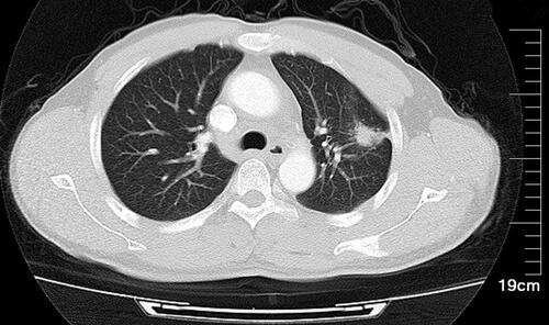
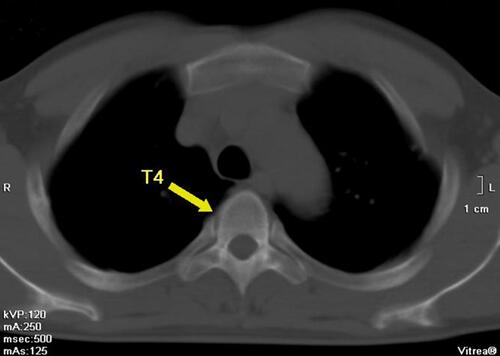
Figure 2 A 16-slice computed tomographic scan revealed a high-density nodule in the fourth thoracic vertebra.
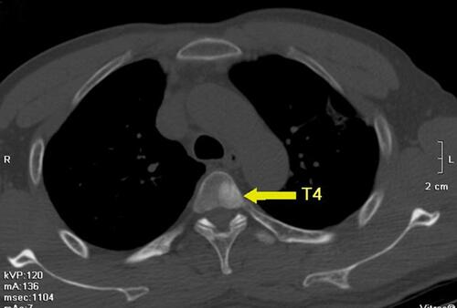
Figure 3 A 99mTc-MDP bone scan revealed bone metastases at the 4th, 5th, 6th, 10th, and 12th thoracic vertebrae and 4th lumbar vertebrae.
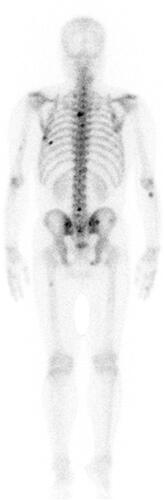
Figure 4 An enlarged lymph node that can be touched on the surface of the body is proven to be structurally abnormal by color ultrasound and is eventually used for pathological biopsy.
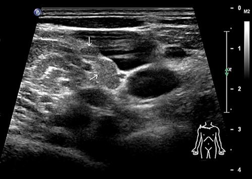
Figure 5 Image of histologic diagnosis using hematoxylin and eosin staining (original ×200), which revealed lung adenocarcinoma metastasis.
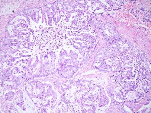
Figure 6 A 16-slice computed tomographic scan revealed that the left lung nodule was significantly smaller than before and was almost absorbed.
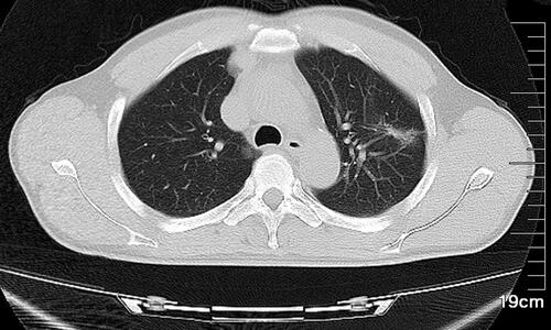
Figure 7 A 99mTc-MDP bone scan revealed that the bone metastases were absorbed.

Figure 8 A 16-slice computed tomographic scan revealed that the high-density nodule in the fourth thoracic vertebra was absorbed.