
Figures & data
Figure 1 Enhanced CT showed that the tumor was located in the anterior mediastinum, presenting a mixed mass of cystic and solid, compressing the right lung (white arrow (A), lung window). The tumor boundary was clear ((B), mediastinal window), with arc-shaped calcification shadows (yellow arrow), solid components (asterisk), and internal septa (white arrow).
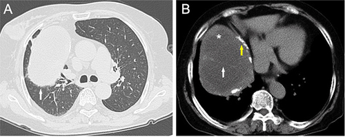
Figure 2 Under the microscope, the mass was multilocular-cystic. The cyst walls varied in different thicknesses, and the solid epithelioid components were like islands (A). The hemorrhage, cholesterol crystal crack, calcification, and edema were in the cyst (B). The epithelioid components were lined in the inner side of some cyst walls, and a small number of lymphocytes were scattered in the epithelioid cell (C). The solid epithelioid areas were surrounded by spindle cell bundles (D). The epithelial cells expressed CK (E) and P63 (F), and the spindle cells expressed Vimentin (G), but the CD20 was negative in both types of cells (H), and the Ki-67 index was less than 5% (I).
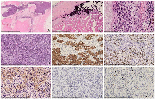
Figure 3 The dual-color separation signal of YAP1 (A) and MAML2 (B) was detected by FISH (the green fluorescent probe was directly hybridized with the distal end of the YAP1 gene, the Orange fluorescent probe was directly hybridized with the proximal end of YAP1 gene; the green fluorescent probe was directly hybridized with the proximal end of MAML2 gene, and the Orange fluorescent probe was directly hybridized with the distal end of MAML2 gene), white arrows indicating separating signals. The YAP1-MAML2 fusion probe detected that there was a yellow fusion signal (C), the Orange probe was 5’ of YAP1 and the green probe was 3’ of MAML2, the yellow arrow indicating the merged signals).

Data Sharing Statement
The datasets supporting the conclusions of this article are included within the article.