Figures & data
Figure 1 (A) A proliferating IH on the cheek of an infant. Note the ulceration in the center of the IH. (B) A proliferating IH is highly cellular with poorly defined vascular spaces (black arrowheads).
Abbreviation: IH, infantile hemangioma.
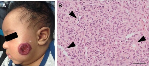
Figure 2 (A) An involuting IH on the arm of a toddler. Note the graying red color when compared to the proliferating IH. (B) Histology: involuting IHs have fewer cells and organized vascular tubular structures (black arrowheads).
Abbreviation: IH, infantile hemangioma.
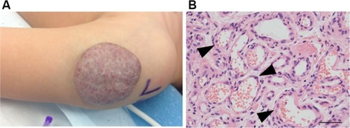
Figure 3 Both proliferating (A) and involuting (B) IH endothelial cells express GLUT1 (green).
Abbreviations: IH, infantile hemangioma; GLUT1, glucose transporter 1.
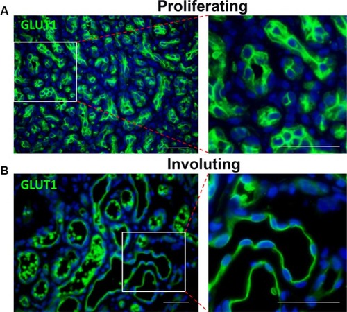
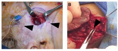
Figure 4 (A) Intraoperative photo demonstrating that IH can be easily separated from the surrounding soft tissue. Note facial monitoring devices in place. These can be used to assist during facial dissection. Course telangiectasia can be seen as well (arrowheads). (B) Feeding vessels to the IH need to be identified (black arrowhead) and isolated before dividing to minimize blood loss.