Figures & data
Figure 1 EEG of a patient with SSPE.
Note: Longitudinal bipolar montage showing periodic slow waves also known as the “Radermecker” complexes.
Abbreviations: EEG, electroencephalogram; SSPE, subacute sclerosing panencephalitis.
Abbreviations: EEG, electroencephalogram; SSPE, subacute sclerosing panencephalitis.
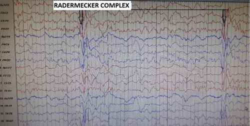
Figure 2 (A–C) The MRI of a 6-year-old girl with SSPE.
Notes: Abnormal signal intensity areas are identified in the periventricular and deep white matter region bilaterally. Abnormal signal are also noted in putamen bilaterally. These areas appear hyperintense on T2-weighted images, hypointense to isointense on T1-weighted images, and there is no evidence of diffusion restriction and no postcontrast enhancement.
Abbreviations: MRI, magnetic resonance imaging; SSPE, subacute sclerosing panencephalitis.
Abbreviations: MRI, magnetic resonance imaging; SSPE, subacute sclerosing panencephalitis.
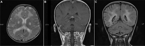
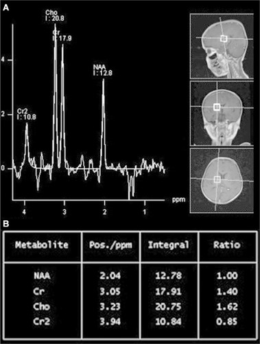
Figure 3 (A and B) The MRS of the 6-year-old girl with SSPE.
Notes: MR spectroscopy shows low NAA levels as well as NAA/Cr and NAA/choline. Patient is the same as seen in .
Abbreviations: MR, magnetic resonance; MRS, magnetic resonance spectroscopy; NAA, nacetyl aspartate; Cr, creatinine.
Abbreviations: MR, magnetic resonance; MRS, magnetic resonance spectroscopy; NAA, nacetyl aspartate; Cr, creatinine.