
Figures & data
Figure 1 Abdominal CT scans. Plain (A) and contrast-enhanced (B) images. Shows hepatic nodules with no calcification with multiple cysts and small daughter cysts communicating hypoechoic mass-like lesion (arrows).
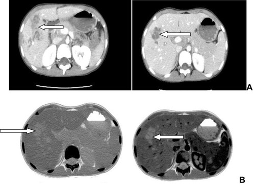
Figure 2 Liver biopsy shows numerous epithelioid granulomas, giant cells, necrosis, and microabscesses and with chronic inflammatory cells, mainly lymphocytes and eosinophils.
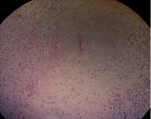
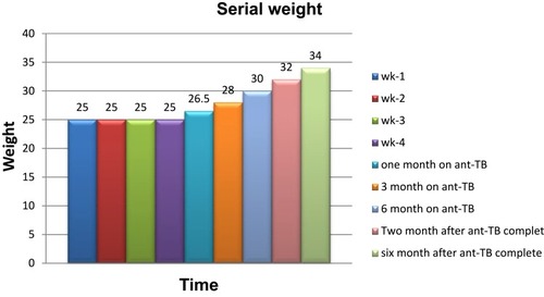
Figure 3 Serial weight measurement during a hospital stay and on follow up after anti-TB treatment began.
Availability of Data and Material
Please contact the correspondence author for data requests.