Figures & data
Figure 1 Cross-section of the mature umbilical cord and schematic representation of mature umbilical cord showing (A) wharton’s jelly with artery, (B) one umbilical vein (single headed arrow), and two arteries (double headed arrow) with Wharton’s jelly H&E. 4x.
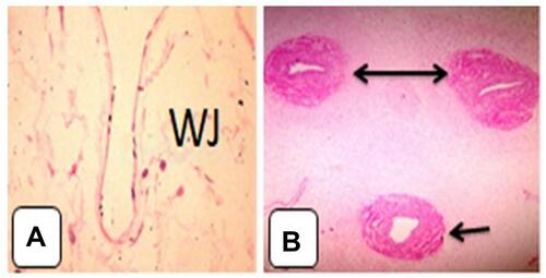
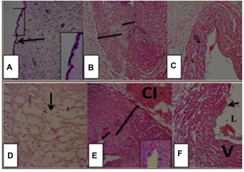
Figure 2 Photomicrographs of umbilical cord sections of control group showing (A) Wharton’s jelly, fibroblast cells and enclosed by amniotic membrane (arrow). The inset is higher magnification of the amniotic membrane. (B) Showing part of umbilical artery with lumen (L), intima (short black line) and thick media (long black line) with no adventitia, surrounded by Wharton’s jelly (lightly stained). (C) Showing the umbilical vein wide lumen, thin intima and thin media, and surrounded by Wharton’s jelly. Umbilical cord sections of diabetic group showing (D) Wharton’s jelly, notice honey combs (↓). (E) Showing part of umbilical artery (A) with narrow lumen (L), intima (short black line) and thick media with cellular debris (long black line), with extra cellular infiltration (CI), with focal erosion of endothelial lining. (F) Showing the wall of umbilical vein (V) with thinner wall and wider lumen (L), with cellular debris in media.