Figures & data
Figure 1 Axial T1-weighted brain MRI demonstrating a well-defined hypointense lesion in the right temporal lobe with significant mass effect and midline shift.
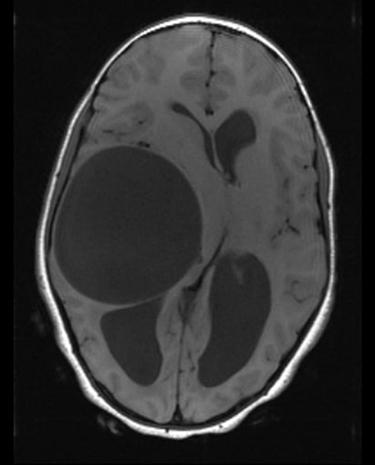
Figure 2 Coronal T2-weighted and axial FLAIR image of the brain demonstrating a well-defined T2 hyperintense lesion that is totally suppressed on the FLAIR sequence, with no sign of perilesional edema.
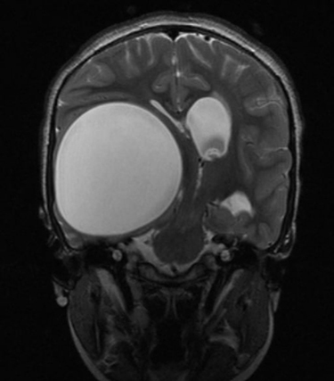
Figure 3 T1-weighted MRI of the brain after gadolinium administration showing a well-defined cystic mass with no enhancement on the cyst wall.
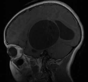
Figure 4 The removed giant intracranial hydatid cyst after the operation.
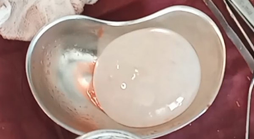
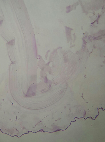
Figure 5 Histopathology microscopic section showing a laminated acellular cyst wall with nucleated germinal layer.