Figures & data
Figure 1 (A) Right eye of an infant who was injected ranibizumab and subsequently lasered. The inadequate laser resulted in persisted aggressive ROP. (B) Three-week follow-up angiography of the same infant who underwent fill-in laser which was performed to address all residual avascular and ischemic regions.
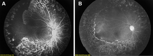
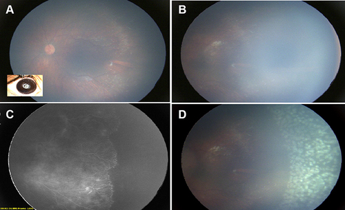
Figure 2 (A) Left eye of a premature infant 11 months after injection of intravitreal bevacizumab who was lost to follow-up. The right eye had a closed funnel retinal detachment. (B) Pre-retinal fibrous tufts were noted in zone 2 with significant persistent peripheral avascular retina. (C) The result was confirmed based on angiography (RetCam 3, Natus, CA, USA). (D) The eye underwent laser photoablation of the avascular retina.