
Figures & data
Figure 1 Stepwise approach for management of sternal cleft.
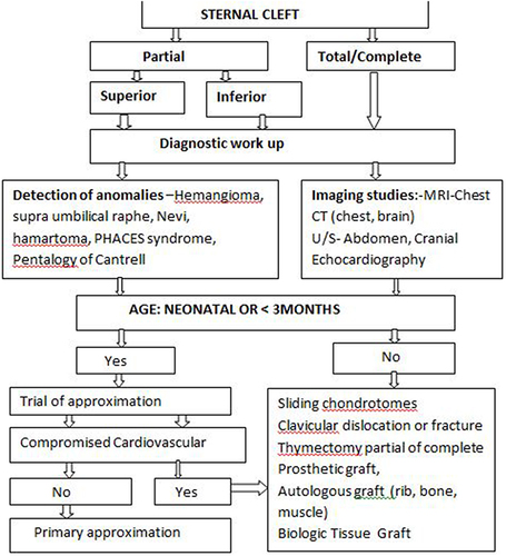
Figure 2 Preoperative: (A) defect on the anterior chest wall with scar-like hypopigmented skin and supraumbilical raphe; (B) marked V-shaped sternal defect.
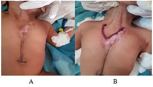
Figure 3 Intraoperative: (A) vertical incision was made, followed by raising skin flap; (B) exposing sternal bars and pectoralis major muscle flaps. The two halves were freed of underlying pleura and pericardium.
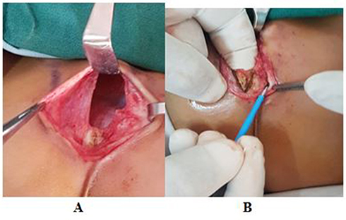
Figure 4 Immediately postprocedure: (A) approximation of sternal bars and muscle flaps done; (B) complication of inward chest retraction is not seen.
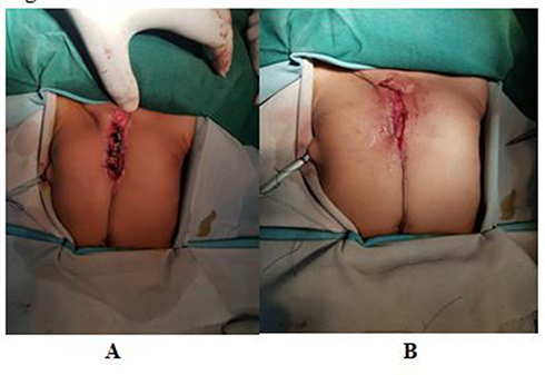
Figure 5 Postoperative: (A) the incision closed layer by layer, cleaned; (B) observed for infection at 2–7 days; (C) at discharge.

Table 1 Summary of seven case reports presented with different types of sternal defect, associated syndromes or anomalies and various techniques of managementCitation2,Citation3,Citation6,Citation8–10