Figures & data
Table 1 Causes of Exocrine Pancreatic Insufficiency in Children
Table 2 Inherited Syndromes Which Causes Exocrine Pancreatic Insufficiency During Infancy:Citation15,Citation21
Table 3 Pancreatic Function Tests
Figure 1 (A) Computed tomography (axial view) of a 8-year-old patient with cystic fibrosis showing pancreatic atrophy and pancreatic calcifications (white arrow). (B) Magnetic resonance cholangiopancreatography (axial view) of an infant with exocrine pancreatic insufficiency from Pearson syndrome showing pancreatic atrophy with fatty replacement (white arrow). (C) Computed tomography (axial view) of a 5-year-old patient with exocrine pancreatic insufficiency from tropical calcific pancreatitis showing extensive pancreatic calcifications (white arrow). (D) Magnetic resonance enterography (coronal view) of a 4-year-old patient with exocrine pancreatic insufficiency showing annular pancreas (white arrow) and pancreatic hypoplasia with small body and tail of the pancreas (black arrow).
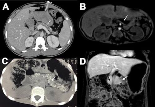
Table 4 Factors to Consider for Poor Response to PERT and Persistence of EPI Symptoms
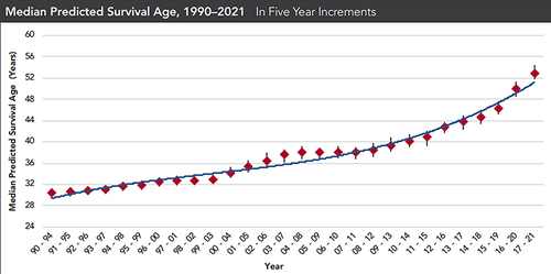
Figure 2 Median predicted survival age of patients with cystic fibrosis from the year 1990 to 2021 in five-year increments (reproduced with permission from the Cystic Fibrosis Foundation).