Figures & data
Figure 1 (A) Left lung hyperinflation and reduced right lung volume along with upper lobe haziness or atelectasis, (B) 12 days later, upper lobe haziness has slightly decreased with an overall decrease in infiltrations along the lung, (C) right upper lobe haziness reappears with widespread lung consolidation and indistinct heart borders.
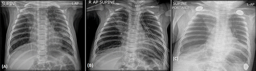
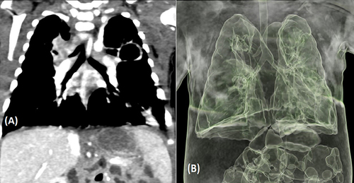
Figure 2 (A) Coronal plane images of chest CT scan demonstrating an extra bronchus origin above the carina, (B) Pulmonary 3D images demonstrating extra bronchus arising from the right lateral aspect of the trachea almost at the level of the carina.