Figures & data
Figure 1 15x15cm hard bifrontal skull mass.
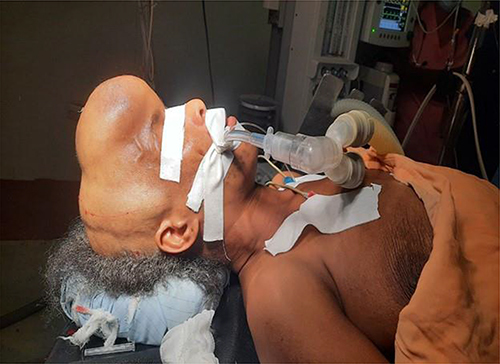
Figure 2 CT scan (the bone window) shows hyperostosis of the frontal bone with a well-defined outline measuring 2.2 cm at its maximum thickness.
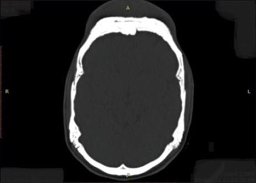
Figure 3 CT scan of the head showing the thickening of the overlying forehead soft tissue and increased paralleling bone lesions.
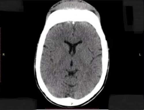
Figure 4 Left lateral view of the CT scan of the head with 3D reconstruction showing the bifrontal mass.
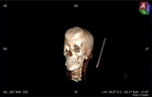
Figure 5 Superior view of the CT scan of the head with 3D reconstruction showing the bifrontal mass.
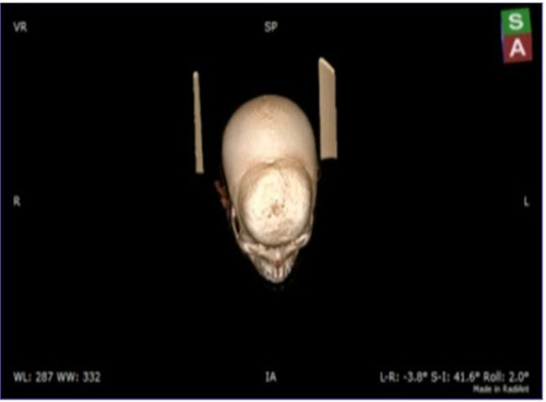
Figure 6 (A and B) Gross pictures showing, a 13 x 12cm slightly irregular Bifrontal calvarial bone with 1.5–3 cm thickness and two separate irregular skin tissues larger measuring 13 x 8cm, smaller measuring 10×2 cm.
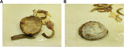
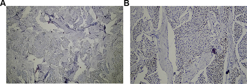
Figure 7 (A and B) Microscopic picture on 10/HPF showing thickened lamellar and woven bone cuffing whorl and syncytium of oval to spindle meningothelial cells.
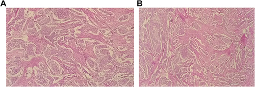
Figure 8 Microscopic picture on 40/HPF showing whorls and syncytium of oval to spindle meningothelial cells.
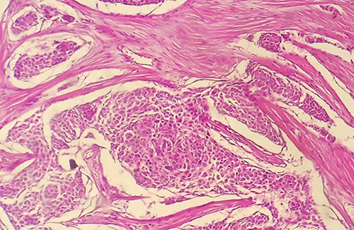
Figure 9 (A and B) Immunostaining for progesterone shows a nuclear pattern of staining.