
Figures & data
Figure 1 Postradiation changes in the chest wall on the right side.
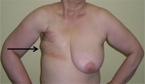
Figure 2 Multichamber pneumatic pump in the form of jacket used in compression therapy.
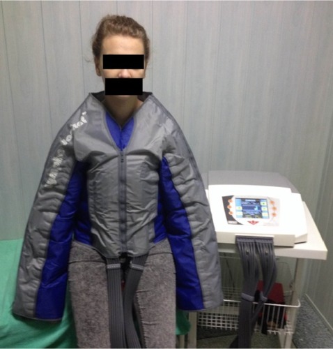
Figure 3 Lymphedema on the operated chest wall in three patients (front view, 3 months after the surgery; back view, 9 months after the surgery; and side view, 6 months after the surgery).
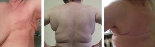
Figure 4 The average age of patients in both subgroups.
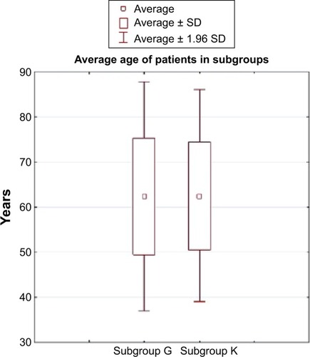
Table 1 The average age of patients in both subgroups
Table 2 Median BMI values in both subgroups
Figure 5 Ultrasound measuring points before mastectomy (measurement I).
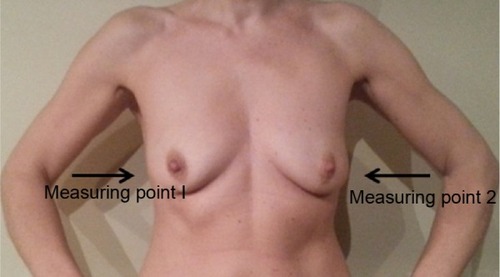
Figure 6 Ultrasound measuring points after mastectomy (measurements II, III, and IV).
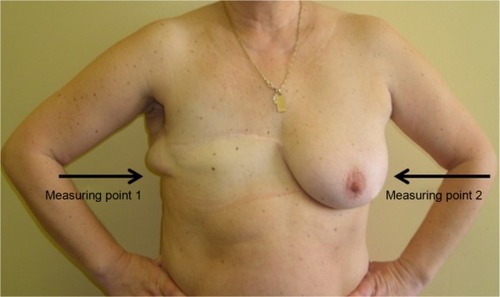
Figure 7 Compression corset which helps relieve lymphatic system (back view, front view, and side view).
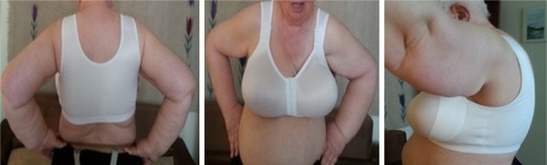
Figure 8 VAS with numeric values.
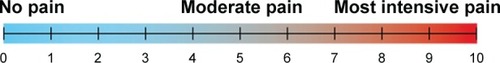
Figure 9 Average thickness ratios of the subcutaneous tissue of the chest wall in subgroup G patients (compression corsets) during the 7-month follow-up.
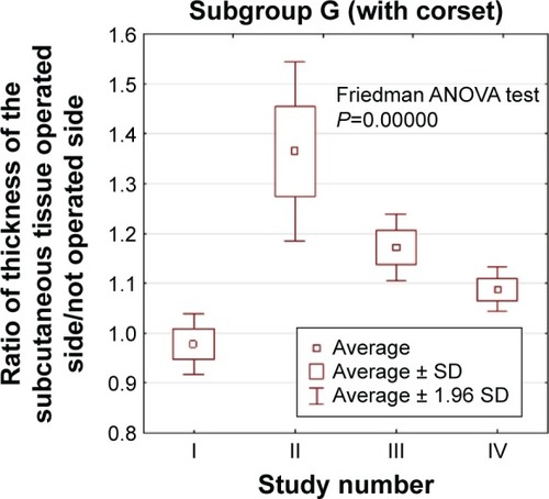
Figure 10 Average thickness ratios of the subcutaneous tissue of the chest wall in subgroup K patients (control) during the 7-month follow-up.
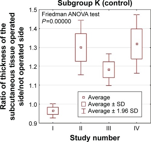
Figure 11 Average thickness ratios of the subcutaneous tissue of the chest wall in both subgroups G (compression corsets) and K (control) during the 7-month follow-up.
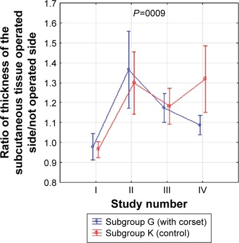
Figure 12 Average thickness ratios of the subcutaneous tissue of the chest wall in subgroup G patients (compression corsets) during the 7-month follow-up with or without additional radiotherapy.

Figure 13 Average thickness ratios of the subcutaneous tissue of the chest wall in subgroup K patients (control subgroup) during the 7-month follow-up with or without additional radiotherapy.
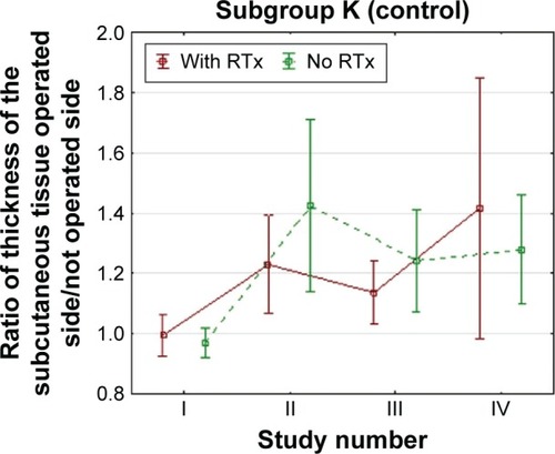
Figure 14 Average thickness ratios of the subcutaneous tissue of the chest wall in both subgroups (G and K) during the 7-month follow-up with or without additional radiotherapy.
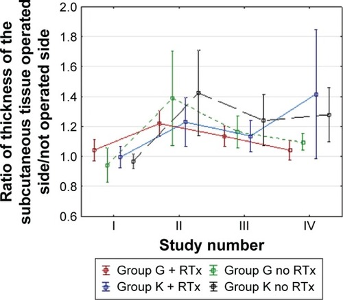
Table 3 The average thickness ratio for the second and fourth measurements in both subgroups with and without additional radiotherapy
Figure 15 Reduction of pain in both subgroups, based on VAS in both subgroups (K, control; G, with corset).
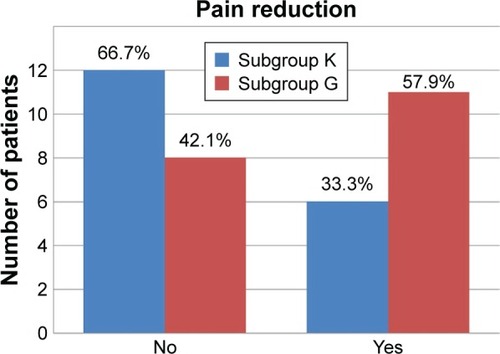
Figure 16 Directions of manual lymph drainage after axillary lymphadenectomy (substitute lymphatic flow areas are depicted in blue and regions of edema and so-called gravity points are in yellow).
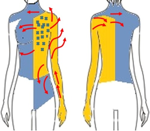
Table 4 Measurement protocol specifying the class of compression of the fabricCitation37