Figures & data
Figure 1 Endoscopic examinations.
Notes: Gastroscopy showed multiple superficial mucosal lesions of duodenum and diffuse mucosal bleeding (A and B), and colonoscopy indicated active oozing of blood from ileocecal junction (C and D).
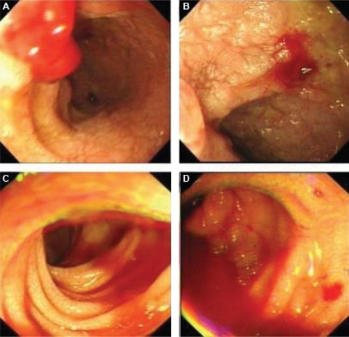
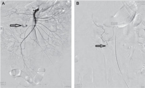
Figure 2 Mesenteric arterial angiography.
Notes: Initial angiography (A) showed a hemorrhage spot in the initial branch of the superior mesenteric artery (indicated by arrow). Selective arterial embolization with gelatin sponge was performed and final angiography (B) demonstrated no signs of arterial bleeding (indicated by arrow).