
Figures & data
Table 1 Differential diagnosis of nail psoriasis and onychomycosis
Figure 1 The thumbs of the patient mainly show nail bed involvement with subungual hyperkeratosis, salmon spot, and onycholysis.
Notes: (A) Before treatment (September 2011). (B) After 3 months of topical treatment with calcipotriol plus betamethoasone dipropionate ointment and clobetasol solution under the nails: the right thumb shows some improvement and the left thum nail has worsened (December 2011).
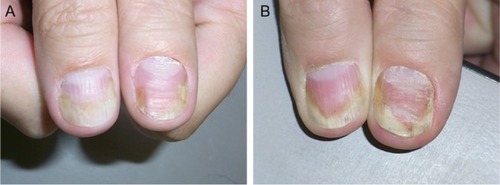
Figure 2 Further development of the nail psoriasis.
Notes: (A) After continued topical treatment (March 2012). (B) After repeated perilesional injections of triamcinolone acetonide crystal suspension (10 mg/mL), a marked improvement is seen (June 2012).
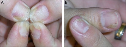
Figure 3 As topical and injection treatments are insufficient and inconvenient, systemic methotrexate is instituted.
Notes: (A) Further improvement of the left thumb nail after 2 months of methotrexate (September 2012). (B) Despite continuous methotrexate therapy, the left thumb nail worsened again (July 2013).
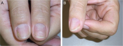
Figure 4 There is residual nail bed psoriasis under methotrexate therapy. Finally, a biological treatment is instituted.
Notes: (A) Slight distal onycholysis and subungual hyperkeratosis (November 2013). (B) Six weeks after the beginning of adalimumab therapy, the patient has 20 clear nails for the first time since >25 years.
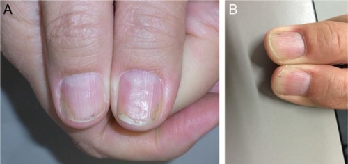
Table 2 Treatment algorithm for nail psoriasis