Figures & data
Figure 1 Chest computed tomography (CT) scan before and after anti-tuberculosis treatment and interventional treatment. (A and B) Segmental infiltration and consolidation in the left lung and apparent stenosis of the left main bronchus before treatment, (C and D) Improvement of left bronchus stenosis and infiltration absorption observed after therapy.
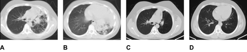
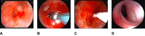
Figure 2 Bronchoscopy images of the left main bronchus stenosis before and after treatment. (A) Scarring stenosis of the left mainstem bronchus, (B) Balloon dilation procedure, (C) Cryoablation post dilation to reduce restenosis, (D) Obvious improvement in the patency of the left mainstem bronchus as shown in post-treatment follow-up examinations.