
Figures & data
Table 1 NODDI findings in Alzheimer’s disease, Parkinson’s disease, multiple sclerosis, and epilepsy
Figure 1 Sample images (A, T1-weighted; B, FLAIR; C, neurite density) of a 48-year-old patient with relapsing-remitting multiple sclerosis. Reduced neurite density can be seen in the demyelinating white matter lesions (arrows).
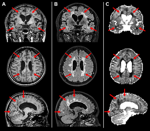
Figure 2 (A) In a 46-year-old patient with focal epilepsy, reduced neurite density is evident in the focal cortical dysplasia (circles). (B) A 17-year-old patient with tuberous sclerosis. Reduced neurite density can be seen in the multiple cortical tubers (arrows). (C) A 37-year-old patient with left temporal lobe epilepsy and hippocampal sclerosis. The abnormal hippocampus shows reduced neurite density and orientation dispersion (arrowheads). (D) A 47-year-old patient with right temporal lobe without any visible lesions on conventional MRI. The right temporal lobe shows reduced neurite density in accordance with the hypometabolic areas of 18F-FDG-PET (broken circles).
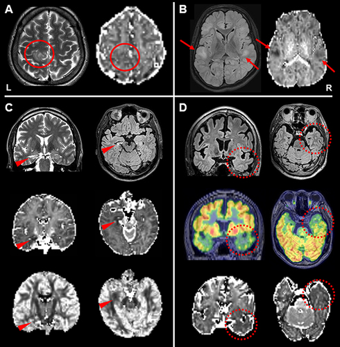
Table 2 NODDI findings in stroke, tumor, and trauma
Table 3 NODDI findings in psychiatry and brain development
Table 4 NODDI findings in other brain diseases and spine