Figures & data
Figure 1 Terminology used to describe the stages of autophagosome assembly during (macro)autophagy.
Notes: (A) Nucleation: the first autophagy proteins begin to assemble at a membrane site that will subsequently mature to become and/or template a single autophagosome. (B) The isolation membrane or phagophore develops: this cytoplasmic sequestering structure is effectively an incomplete/unsealed autophagosome which grows, is shaped, and ultimately seals to form a 3D structure with double membrane (thereby engulfing parts of the cytoplasm and/or cargo) that is the completed autophagosome (C). The autophagosome is a motile transport vacuole that leaves the site of nucleation/assembly (which in mammalian cells is rapidly dismantled) and traffics to the lysosome (D), where it fuses to form an autolysosome (E). Here, macromolecules are degraded into their constitutive building blocks by acidic hydrolases that are released back into the cytoplasm via specific retrograde pumps (E).
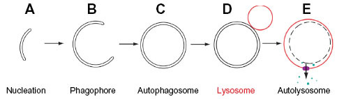
Figure 2 Protein complexes acting during starvation-induced autophagosome assembly.
Notes: (A) mTorC1 and its accessory partners. (B) The ULK1 complex and its constitutive components. (C) The autophagy PI(3) kinase complexes with autophagy activating and inhibiting actions. The core complex is shown in orange and red. (D) The conjugation apparatus that acts to form the ATG5-12 complex. (E) ATG proteins required for LC3 lipidation (a process that requires ATG5-12-16 targeting to the site of autophagosome nucleation).
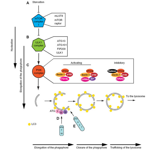
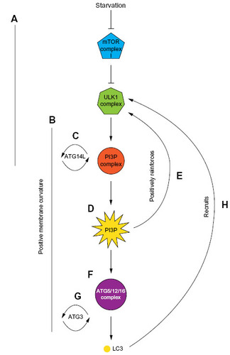
Figure 3 Positive reinforcement of the hierarchical recruitment of autophagy proteins during autophagosome assembly.
Notes: (A) Early stages of autophagosome assembly. (B) Positive membrane curvature at the autophagosome assembly site. (C) ATG14L has affinity for curved membranes. (D) The VPS34 PI(3)kinase complex triggers localized PI3P formation. (E) This platform of PI3P stabilizes the ULK1 complex at the phagophore through a PI3)-binding region in ULK1. (F) The ATG5/12/16 complex is recruited to PI3P positive phagophores and WIPI2b. (G) ATG3 binds to and lipidates LC3 at curved membranes. (H) LC3 stabilizes/recruits the ULK1 complex due to a LIR motif in ULK1.