Figures & data
Figure 1 Schematic representation of the phases and timeline of the skin healing process in mice. Four overlapping phases shown schematically result in wound closure. The cellular processes at each stage are described in detail in “Normal skin healing process in mice”.
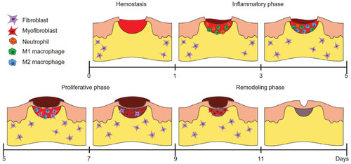
Table 1 Influence of genetic deletion of matricellular proteins on skin healing in mice
Figure 2 α-Smooth muscle actin expression is significantly reduced in periostin knockout mice compared with wild-types at 7 days postwounding. Red arrows mark the wound edge.
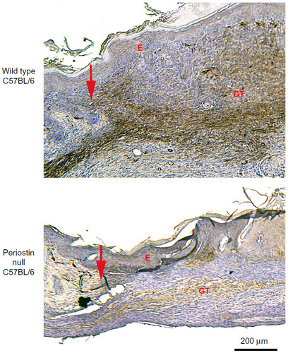
Table 2 Known expression profiles of matricellular proteins in human wounds
Figure 3 Periostin expression is reduced in human nonhealing skin wounds and correlates with a reduction in α-SMA expression and collagen content. Tissue was isolated at elective amputation from an 83-year-old female with type 2 diabetes.
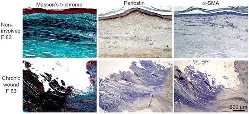
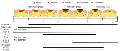
Figure 4 Temporal expression profiles of matricellular proteins during skin healing.