Figures & data
Figure 1 Schematic diagram of exosome biogenesis and secretion.
Notes: Exosomes are generated in the endosomal structure and secreted via endosomal pathways. The exosomes contain specific proteins and microribonucleic acid, mediating intercellular communication to modify the different biological function of target cells.
Abbreviations: EVs, extracellular vesicles; ESCRT, endosomal sorting complex required for transport.
Abbreviations: EVs, extracellular vesicles; ESCRT, endosomal sorting complex required for transport.
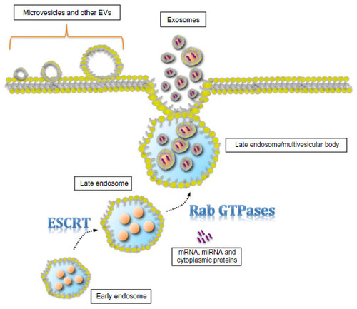
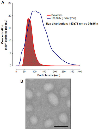
Figure 2 Extracellular vesicle (EV) size distribution and exosome morphology.
Notes: (A) Representative vesicle size distribution measured by the nanoparticle tracking analysis NanoSight NS500 instrument. Blue: 100,000× g pellet (EVs); red: enriched exosome population. (B) Representative electron micrograph of exosome population, scale bar 100 nm.