Figures & data
Figure 1 Schematic representation of different domains of BoNT/A.
Note: The heavy chain receptor binding domain is marked in red, green is the heavy chain translocation domain, and the light chain catalytic domain is colored blue.
Abbreviation: BoNT/A, botulinum neurotoxin type A.
Abbreviation: BoNT/A, botulinum neurotoxin type A.
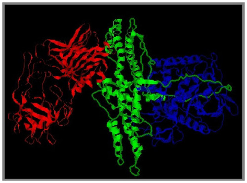
Figure 2 Schematic model of mode of action of botulinum neurotoxins.
Notes: (A) Synaptic vesicles containing neurotransmitters dock and fuse with the plasma membrane through interaction of the SNARE proteins (Synaptobrevin, SNAP-25, and Syntaxin). (B) Botulinum neurotoxin binds to the presynaptic membrane through gangliosides and a protein receptor followed by internalization into the endosomes via endocytosis. Following this, the light chain is translocated across the membrane into the cytosol where it acts as a specific endopeptidase against either of the SNARE proteins. BoNTs cleave their substrates before the formation of SNARE complex. Copyright © 2009. Caister Academic Press. Reproduced from Kukreja R, Singh BR. Botulinum neurotoxins-structure and mechanism of action. In: Proft T, editor. Microbial Toxins: Current Research and Future Trends. Norfolk: Caister Academic Press; 2009:15–40.Citation75
Abbreviations: SNARE, sensitive factor attachment protein receptor; BoNTs, botulinum neurotoxins.
Abbreviations: SNARE, sensitive factor attachment protein receptor; BoNTs, botulinum neurotoxins.
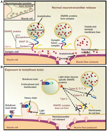
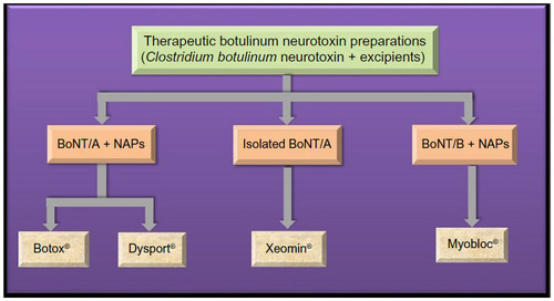
Figure 3 Contents of commercially available therapeutic botulinum formulations.
Abbreviations: BoNT/A, botulinum neurotoxin type A; NAPs, neurotoxin-associated proteins; BoNT/B, botulinum neurotoxin type B.