
Figures & data
Table 1 Classification of aaRSs
Figure 1 Schematic representation of localization and function of HctPheRS and HmtPheRS.
Abbreviations: FARS, HctPheRS encoding gene; FARS2, HmtPheRS encoding gene; HctPheRS, human cytosolic phenylalanyl-tRNA synthetase; HmtPheRS, human mitochondrial phenylalanyl-tRNA synthetase; Phe, phenylalanine; Tyr, tyrosine.
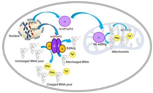
Table 2 Structural organisation of eubacterial PheRS
Table 3 Domain organisation divergence between the kingdoms
Figure 2 Crystal structures of HctPheRS and ttPheRS.
Notes: (A) Adapted from Structure; 18(3); Finarov I, Moor N, Kessler N, Klipcan L, Safro MG; Structure of human cytosolic phenylalanyl-tRNA synthetase: evidence for kingdom-specific design of the active sites and tRNA binding patterns; 343–353; Copyright © 2010; with permission from Elsevier.Citation27 Crystal structure of HctPheRS (PDB ID: 3L4G). Domain architecture of the two αβ heterodimers are shown with one heterodimer lacking the N-terminal domain of the α subunit. α subunit: N-terminal domain containing DBD-1, DBD-2, and DBD-3 colored green, the linker region colored yellow, catalytic domains A1 and A2 colored red and blue, respectively. β subunit: B1, B3, B4 domains colored grey, B5 domain colored brown, B6 domain colored purple, and the B7 domain colored cyan. (B) Adapted from Moor N, Kotik-Kogan O, Tworowski D, Sukhanova M, Safro M. The crystal structure of the ternary complex of phenylalanyl-tRNA synthetase with tRNAPhe and a phenylalanyl-adenylate analogue reveals a conformational switch of the CCA end. Biochemistry. 2006; 45(35):10572–10583.Citation33 Crystal structure of ttPheRS complexed with tRNAPhe and PheOH-AMP (PDB ID: 2iy5). Domain architecture of one αβ heterodimer is shown with N-terminal coiled coil of α subunit colored green, catalytic domains A1 and A2 colored red and blue, respectively, β subunit domain B1 colored grey, B2 domain colored pink, B3 domain colored orange, B4 domain colored olive green, B5 domain colored brown, B6 domain colored purple, B7 domain colored cyan, and B8 domain colored yellow.
Abbreviations: DBD, DNA binding domain; HctPheRS, human cytosolic phenylalanyl-tRNA synthetase; ttPheRS, Thermus thermophiles phenylalanyl-tRNA synthetase; PDB, Protein Data Bank; PheOH-AMP, phenylalaninyl-adenylate.
Abbreviations: DBD, DNA binding domain; HctPheRS, human cytosolic phenylalanyl-tRNA synthetase; ttPheRS, Thermus thermophiles phenylalanyl-tRNA synthetase; PDB, Protein Data Bank; PheOH-AMP, phenylalaninyl-adenylate.
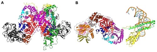
Figure 3 Crystal structures of HmtPheRS.
Notes: (A) Adapted from Structure; 16(7); Klipcan L, Levin I, Kessler N, Moor N, Finarov I, Safro M; The tRNA-induced conformational activation of human mitochondrial phenylalanyl-tRNA synthetase; 1095–1104; Copyright © 2008; with permission from Elsevier.Citation47 Crystal structure of HmtPheRS (PDB ID: 3CMQ).Citation47 (B) Adapted from J Mol Biol; 415(3); Klipcan L, Moor N, Finarov I, Kessler N, Sukhanova M, Safro MG; Crystal structure of human mitochondrial PheRS complexed with tRNA(Phe) in the active “open” state; 527–537; Copyright © 2012; with permission from Elsevier.Citation48 Crystal structure of HmtPheRS complexed with tRNAPhe (PDB ID: 3TUP).Citation48 Domain architecture of HmtPheRS with the N-terminal domain colored green, the catalytic domain colored red, the linker region colored purple, and the anticodon binding domain colored yellow.
Abbreviation: HmtPheRS, human mitochondrial phenylalanyl-tRNA synthetase.
Abbreviation: HmtPheRS, human mitochondrial phenylalanyl-tRNA synthetase.
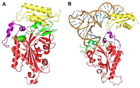
Table 4 Disease-related mutations of HmtPheRS
Figure 4 Sites of genetic mutations in HmtPheRS.
Abbreviations: HmtPheRS, human mitochondrial phenylalanyl-tRNA synthetase; MTS, mitochondrial targeting sequence.
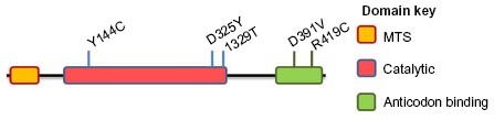
Table 5 Antisynthetase antibodies