Figures & data
Figure 1 Electrocardiography: shows left ventricular hypertrophy and left bundle branch block.
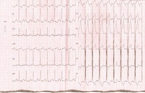
Figure 2 PA chest X-ray shows enlargement of cardiac shadow.
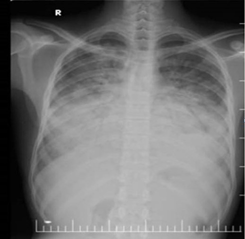
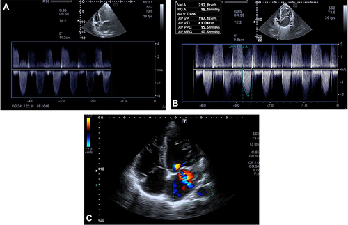
Figure 3 (A–C) Echocardiographic findings: five-chamber view of two-dimensional echocardiography obtained at the time of presentation revealed noncompaction layers of left ventricular with subaortic membranous senosis and four chamber view with Doppler.