Figures & data
Figure 1 Pulmonary vein ablation catheter electrograms from the left inferior pulmonary vein.
Notes: (A) Conductance pattern before ablation; (B) entrance block demonstrated after successful ablation with pulmonary vein ablation catheter; (C) during stimulation before ablation; and (D) exit block demonstrated after successful ablation with pulmonary vein ablation catheter.
Abbreviations: CS, coronary sinus electrode; PVAC, pulmonary vein ablation catheter.
Abbreviations: CS, coronary sinus electrode; PVAC, pulmonary vein ablation catheter.
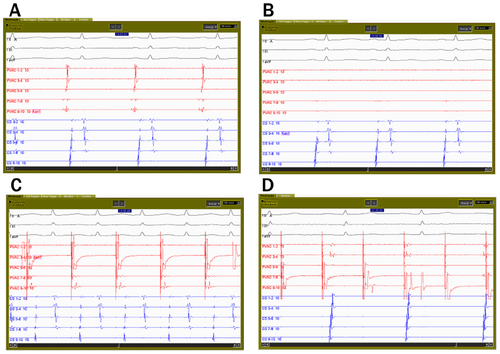
Table 1 Baseline characteristics
Table 2 Procedural data
Table 3 Univariate analysis of predictors of long-term freedom from ablation
Figure 2 Percent of patients free from AF at different time points during follow-up.
Notes: Blue bars indicate patients treated with the PVAC; grey bars are patients treated with NavX-guided point-by-point ablation. (A) All patients; (B) patients with paroxysmal AF; and (C) patients with persistent/long-lasting persistent AF.
Abbreviations: PVAC, pulmonary vein ablation catheter; AF, atrial fibrillation; n, number.
Abbreviations: PVAC, pulmonary vein ablation catheter; AF, atrial fibrillation; n, number.
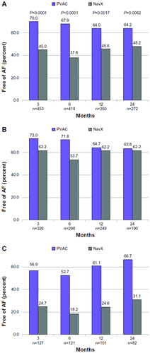
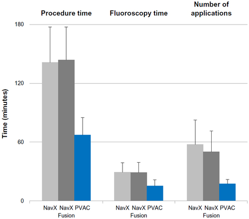
Figure 3 Procedure and fluoroscopy times, and number of applications for ablation guided by NavX and NavX Fusion, as well as PVAC ablation, respectively.
Notes: Lines indicate SD. Differences were significant for all comparisons between PVAC and NavX or NavX Fusion. No significant differences were found between NavX and NavX Fusion.
Abbreviations: PVAC, pulmonary vein ablation catheter; SD, standard deviation.
Abbreviations: PVAC, pulmonary vein ablation catheter; SD, standard deviation.