Figures & data
Figure 1 Schematic representation and photograph of the HIFU balloon.
Notes: (A) The noncompliant balloon, optimal for pulmonary vein ablation, is attached to a steerable catheter. An ultrasound crystal is located at the proximal part of the balloon. The balloon is filled with carbon dioxide and forms an interface allowing the reflection of the ultrasound beam. The acoustic power of the balloon is 45 W. (B) The HIFU catheter with the inflated balloon. Reproduced from Schmidt B, Chun KRJ, Kuck KH, Antz M. Pulmonary vein isolation by high intensity focused ultrasound. Indian Pacing and Electrophysiology Journal. 2006;7(2):126–133.Citation32 Copyright © 2007 Schmidt et al.
Abbreviation: HIFU, high-intensity focused ultrasound.
Abbreviation: HIFU, high-intensity focused ultrasound.
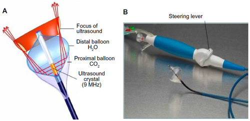
Figure 2 Lesion formation with HIFU.
Notes: HIFU lesion is shown in a model of a canine heart. (A) A deep, septal lesion is shown. Arrow denotes the deepness of the HIFU lesion. (B) A discrete necrotic lesion is shown with sparing of the surrounding tissue. Reprinted from the Journal of the American Society of Echocardiography; 2007;20; Otsuka R, Fujikura K, Abe Y, et al; Extracardiac ablation of the left ventricular septum in beating canine hearts using high-intensity focused ultrasound; 1400–1406; Copyright © 2007 American Society of Echocardiography; Published by Elsevier; All rights reserved; with permission from Elsevier.Citation32
Abbreviations: HIFU, high-intensity focused ultrasound; RV, right ventricle; LV, left ventricle.
Abbreviations: HIFU, high-intensity focused ultrasound; RV, right ventricle; LV, left ventricle.
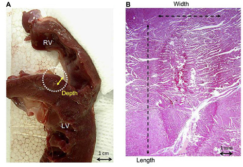
Figure 3 Location of structures prone to injury during balloon-based AF ablation.
Notes: Schematic image demonstrating anatomical relationship between the phrenic nerve and esophagus in relation to the pulmonary vein ostium. (A) Top, anteroposterior view and (B) top view.
Abbreviations: AF, atrial fibrillation; Ao, descending aorta; Eso, esophagus; HIFU, high-intensity focused ultrasound; LA, left atrium; RSPV, right superior pulmonary vein; RT PN, right phrenic nerve; SVC, superior vena cava.
Abbreviations: AF, atrial fibrillation; Ao, descending aorta; Eso, esophagus; HIFU, high-intensity focused ultrasound; LA, left atrium; RSPV, right superior pulmonary vein; RT PN, right phrenic nerve; SVC, superior vena cava.
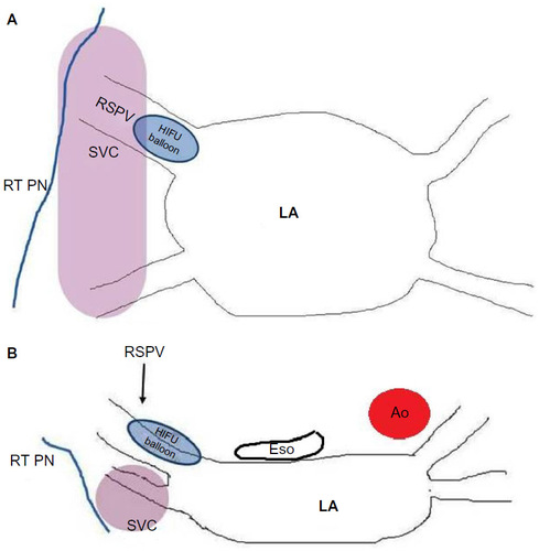
Figure 4 The Epicor epicardial HIFU system.
Notes: The epicardial HIFU system is placed around the four pulmonary veins to create the “box” lesion at the time of a cardiac surgical procedure for AF ablation. St Jude Medical is a trademark of St. Jude Medical, Inc. and its related companies. Reprinted with permission of St. Jude Medical, © 2015. All rights reserved.
Abbreviations: HIFU, high-intensity focused ultrasound; AF, atrial fibrillation.
Abbreviations: HIFU, high-intensity focused ultrasound; AF, atrial fibrillation.
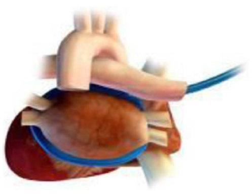
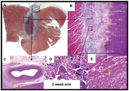
Figure 5 Epicardial ventricular ablation over a coronary artery.
Notes: (A) HIFU application on the epicardial aspect of the left ventricle over the left anterior descending artery. Note the absence of coronary artery injury. (B) Image (4×, Masson’s trichrome [MA]) shows transition from viable myocytes (left) to fibrotic border with inflammation (middle) to necrotic myocytes (right); (C) image (4×, hematoxylin-eosin [HE]) shows LAD with adventitial fibrosis (yellow arrow); (D) image (40×, HE) shows calcification of necrotic myocytes with giant cell (yellow arrow); (E) image (40×, HE) shows necrotic myocytes with nuclear loss (yellow arrow). Adapted from Koruth JS, Dukkipati S, Carrillo RG, et al. Safety and efficacy of high-intensity focused ultrasound atop coronary arteries during epicardial catheter ablation. Journal of Cardiovascular Electrophysiology. 2011;22:1274–1280.Citation31 Copyright © 2011 Wiley Periodicals, Inc.
Abbreviations: HIFU, high-intensity focused ultrasound; LAD, left anterior descending artery.
Abbreviations: HIFU, high-intensity focused ultrasound; LAD, left anterior descending artery.