
Figures & data
Figure 1 Male sex: ultrasound identification of the male sex at the first trimester ultrasound scan shows tubercle’s angle >30°.
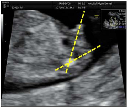
Figure 2 Female sex: ultrasound identification of the female fetal sex at first trimester ultrasound scan shows the genital tubercle parallel to the spinal column.
Note: Inset shows full ultrasound scan.
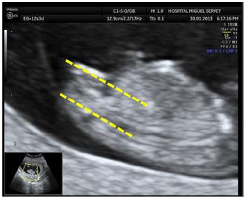
Figure 3 Success rate according to fetal sex algorithm.
Abbreviation: CRL, crown–rump length.
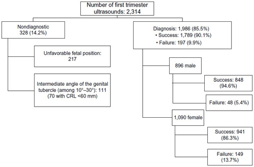
Figure 4 Global success rate according to CRL and divided by sex.
Abbreviation: CRL, crown–rump length.
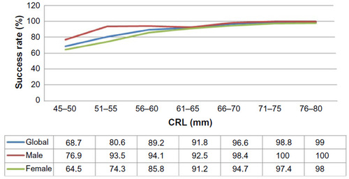
Table 1 Results of univariate and multivariate analysis of independent variables and its relation to the success rate in the diagnosis of fetal sex