
Figures & data
Table 1 Causes of infantile erythroderma
Figure 1 Infant with a metabolic disorder, widespread hyperkeratotic scale, but no collodion is present.
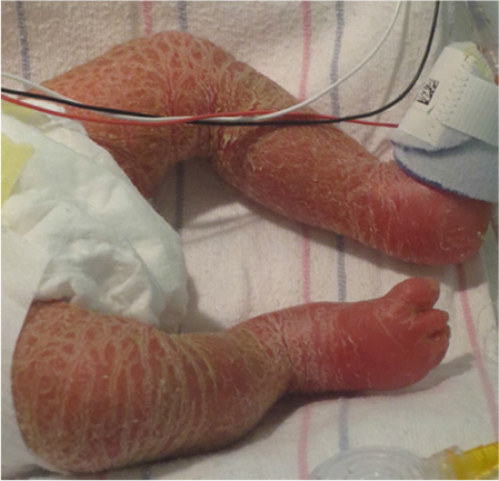
Figure 2 Neonate with Netherton syndrome.
Note: Skin is diffusely erythematous with fine scaling.
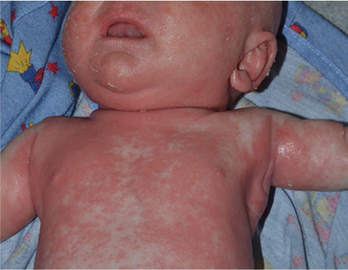
Figure 3 Psoriasis in infants often localizes to the diaper area.
Note: Periumbilical involvement is also common. Plaques are well demarcated.
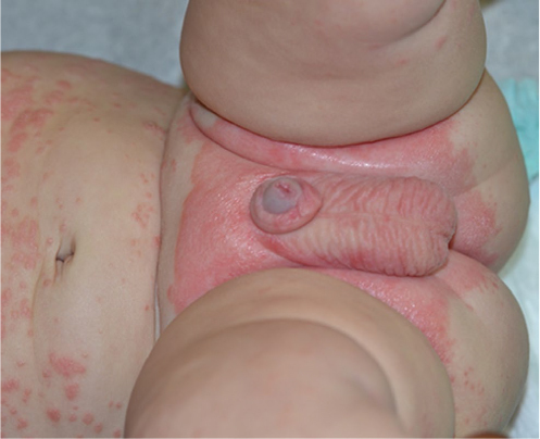
Figure 4 A collodion membrane encases this infant’s body, including his feet and toes.
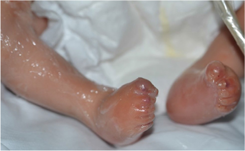
Figure 5 Algorithm for diagnosis of the erythrodermic infant.
Note: *Ichthyoses include congenital ichthyosiform erythroderma, lamellar ichthyosis, harlequin ichthyosis, and rarely Sjogren–Larsson, trichothiodystrophy, and neutral lipid storage disease.
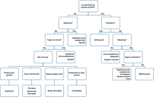
Table 2 Initial diagnostic testing for the erythrodermic infant