Figures & data
Figure 1 Abdominal ultrasound showing type I choledochal cyst.
Notes: Abdominal sonogram transverse view of pancreas. Arrow points to dilated bile duct measuring 4.3 mm.
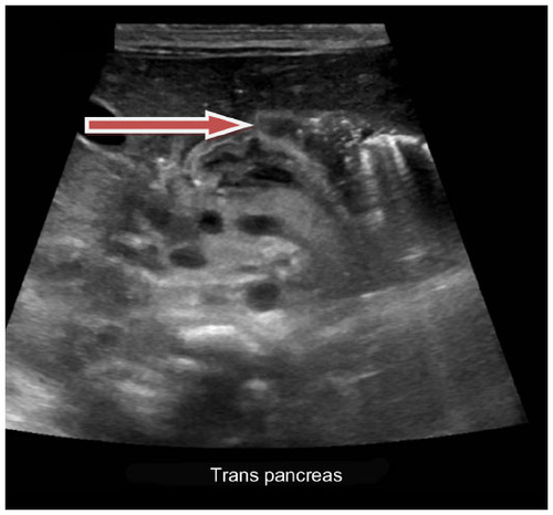
Figure 2 MRCP for better visualization of the choledochal cyst.
Note: MRCP revealed choledochal cyst (arrow) with extension involving distal aspects of right and left hepatic ducts.
Abbreviation: MRCP, magnetic resonance cholangiopancreatography; F, front; L, left; P, posterior.
Abbreviation: MRCP, magnetic resonance cholangiopancreatography; F, front; L, left; P, posterior.
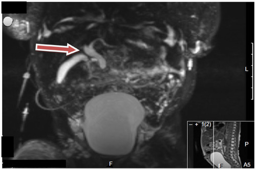
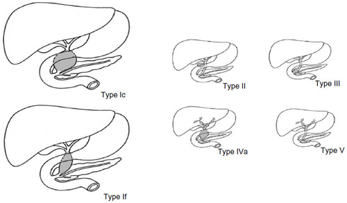
Figure 3 Todani classification of choledochal cysts.
Note: Reprinted from The American Journal of Surgery, 134(2), Todani T, Watanabe Y, Narusue M, Tabuchi K, Okajima K, Congenital bile duct cysts: Classification, operative procedures, and review of thirty-seven cases including cancer arising from choledochal cyst, 263–269, Copyright © (1977), with permission from Elsevier.Citation19
Abbreviations: Type I, cystic (Ic) or fusiform (If); Type II, diverticulum; Type III, choledochocele; Type IV, multiple extrahepatic and intrahepatic duct cysts (IVa) or multiple extrahepatic duct cysts (IVb); Type V, intrahepatic duct cysts (single or multiple).
Abbreviations: Type I, cystic (Ic) or fusiform (If); Type II, diverticulum; Type III, choledochocele; Type IV, multiple extrahepatic and intrahepatic duct cysts (IVa) or multiple extrahepatic duct cysts (IVb); Type V, intrahepatic duct cysts (single or multiple).