Figures & data
Figures 1–8 Evidence of WEEV infection in Cx. tarsalis tissues. 1a) Infected cardia and anterior midgut, HVP strain, 5-log group, 21 days postinfective blood meal. 1b) Cardia and anterior midgut, negative control. 2a) Infected brain and subesophageal ganglion, HVP strain, 5-log group, 21 days postinfective blood meal. 2b) Brain and subesophageal ganglion, negative control. 3a) Infected thoracic ganglion, HVP strain, 5-log group, 21 days postinfective blood meal. 3b) Thoracic ganglion, negative control. 4a) Infected salivary glands, HVP strain, 5-log group, 21 days postinfective blood meal. 4b) Salivary glands, negative control. 5a) Infected eggs, COAV strain, 5-log group, 21 days postinfective blood meal. 5b) Eggs, negative control. 6a) Infected abdominal ganglion, HVP strain, 3-log group, 7 days postinfective blood meal. 6b) Abdominal ganglion, negative control. 7a) Infected retrocerebral complex, HVP strain, 5-log group, 7 days postinfective blood meal. 7b) Retrocerebral complex, negative control. 8a) Infected trachea, KNWR strain, 5-log group, 14 days postinfective blood meal. 8b) Trachea, negative control. In all figures, rusty-brown staining in internal tissues corresponds to positive ABC reactions, indicating the presence of WEE viral antigen. Notice that the cuticle (exoskeleton) often displays a similar brown coloring (eg, in ), which is not indicative of positive ABC reactions.
Figure 9 Infection, dissemination, and salivary gland infection rates among experimental individuals.
Table 1 Overall rates of infection, dissemination, and salivary gland infection
Figure 10 Rates of dissemination and cardia infection among infected individuals. A significant correlation (P < 0.05) between these variables was found in the HVP strain, 5-log group. No significant correlations were found in the 3-log group. Asterisks mark time points where no infected specimens were found.
Figure 11 Infection rates of neural tissue and salivary glands among infected individuals. A significant correlation (P < 0.05) between these variables was found in the 5-log group in all strains except the HVP. No significant correlations were found in the 3-log group. Asterisks mark time points where no infected specimens were found.
Figure 12 Midgut infection barrier (MIB) rates. Ingestion of 5-log of virus is associated with a significant decrease (P < 0.05) in the percentage of individuals displaying a MIB in all strains. In the HVP strain, MIB was completely overwhelmed in the 5-log group.
Table 2 Overall rates of individuals displaying a midgut infection barrier, midgut escape barrier, and salivary gland infection barrier
Figure 13 Midgut escape barrier (MEB) rates. No significant difference between dose groups was found in overall MEB rates for any strain. Asterisks mark time points where MEB rates could not be estimated because no indication of gut infection was seen (MIB rate = 100%).
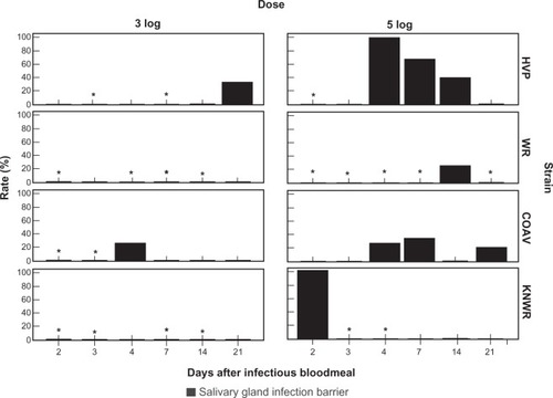
Figure 14 Salivary gland infection barrier (SIB) rates. No significant differences were found either between dose groups, or between strains. Asterisks mark time points where rates could not be estimated due to the absence of individuals with disseminated infections.