Figures & data
Figure 1 (A, B) RVFV antigen in chorionated eggs showing pathology and RVFV antigen-negative germarium (B). (C) Chorionated egg with distinct traces of RVFV antigen. (D) Uninfected chorionated egg with antigen-positive ovariolar sheath. (E) Uninfected chorionated eggs with RVFV antigen-positive ovarian sheath. (F) Sagittal section in posterior part of the abdomen showing antigen-positive oviduct epithelium and tissues in proximity to the spermathecae. (G) Antigen-positive small tracheae associated with the ovaries. (B) Section of spermathecae and adjacent antigen-positive tissues, and antigen-negative accessory gland. (I–L) Negative control sections.
Note: Scale lines (µm): A–E, 150; F, 100; G, 20; H, 50; I–L, 50.
Abbreviations: Ag, RVFV antigen; AG, accessory gland; Ch, chorion; CO, common oviduct; FE, follicular epithelium; Ge, germarium; OS, ovary sheath; OvS, ovariole sheath; RVFV, Rift Valley fever virus; Sp, spermatheca; TL, tracheal lumen; Tr, trachea; UE, uninfected egg; UF, uninfected follicle; Yo, yolk.
Abbreviations: Ag, RVFV antigen; AG, accessory gland; Ch, chorion; CO, common oviduct; FE, follicular epithelium; Ge, germarium; OS, ovary sheath; OvS, ovariole sheath; RVFV, Rift Valley fever virus; Sp, spermatheca; TL, tracheal lumen; Tr, trachea; UE, uninfected egg; UF, uninfected follicle; Yo, yolk.
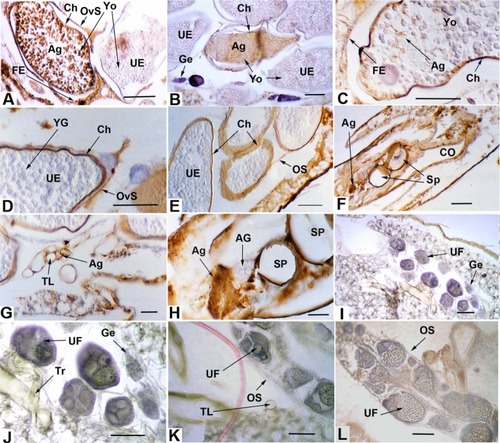
Table 1 RVFV antigen in Aedes mcintoshi female reproductive tissues