Figures & data
Figure 1 Fluorescence microscopy showing dendrites (A and B) and axons (C and D) of layer V pyramidal neurons in the cerebral cortex of mock-infected (A and C) and moribund challenge virus standard-infected (B, D, and D inset) yellow fluorescent protein mice.Citation43 In infected mice, beading is observed in a minority of dendrites (B), and more axons are involved (D). There are no abnormalities in the dendrites (A) or axons (C) of mock-infected mice. Axons in mock-infected mice are slightly varicosed (C), which is characteristic of these fibers. Fluorescence microscopy shows rabies virus antigen (red) in the perikaryon and dendrite of a yellow fluorescent protein-expressing neuron (D inset). Morphology of the cerebellar mossy fibers of mock-infected (E) and moribund challenge virus standard-infected yellow fluorescent protein mice (F). Mossy fiber axons in the cerebellar commissure of moribund mice show severe beading (F), whereas no abnormalities were observed in mock-infected mice (E). Axons in the inferior cerebellar peduncles are normal in mock-infected mice (G) and show marked beading in challenge virus standard-infected moribund mice (H). A–D) ×230; D) inset, ×220; E, F) ×80; G, H) ×350.
Copyright © 2010, American Society for Microbiology. Adapted with permission from Scott CA, Rossiter JP, Andrew RD, Jackson AC. Structural abnormalities in neurons are sufficient to explain the clinical disease and fatal outcome in experimental rabies in yellow fluorescent protein-expressing transgenic mice. J Virol. 2008;82:513–521.Citation43
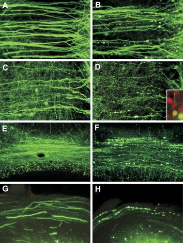
Figure 2 Challenge virus standard infection, but not mock infection, induces formation of 4-hydroxy-2-nonenal (4-HNE)-labeled axonal swellings.Citation51 Fluorescence microscopy showing mock-infected (A, C, and E) and challenge virus standard-infected (B, D, and F) dorsal root ganglion neurons at 72 hours postinfection. β-tubulin (A and B) is a marker of dorsal root ganglion neuronal cell bodies and axons (red) and expression of β-tubulin in challenge virus standard-infected neurons (B) showed multiple axonal swellings but a lack of axonal swellings in mock-infected neurons (A). 4-HNE (green) was poorly expressed in the axons of mock-infected dorsal root ganglion neurons (C) but showed greater expression in the axons of challenge virus standard-infected neurons (D) and accumulation in regions with axonal swellings (D). In challenge virus standard-infected neurons, merging of signals for β-tubulin and 4-HNE (yellow) showed there was strong expression of these elements in axons, both with axonal swellings (arrowheads) and without axonal swellings (arrow, F), but not in mock infected neurons (E).
Copyright © 2010, American Society for Microbiology. Adapted with permission from Jackson AC, Kammouni W, Zherebitskaya E, Fernyhough P. Role of oxidative stress in rabies virus infection of adult mouse dorsal root ganglion neurons. J Virol. 2010;84:4697–4705.Citation51
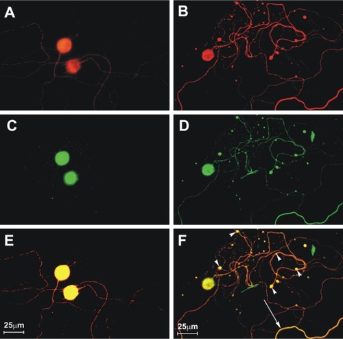
Table 1 Indigenously acquired cases of human rabies from bats in the US and Canada (1950–2009)Table Footnote*
Figure 3 Distribution of the major rabies virus variants among wild terrestrial reservoirs in the US and Puerto Rico in 2008 (from Centers for Disease Control and Prevention [USA]).Citation64
![Figure 3 Distribution of the major rabies virus variants among wild terrestrial reservoirs in the US and Puerto Rico in 2008 (from Centers for Disease Control and Prevention [USA]).Citation64](/cms/asset/e0056eb9-40a1-4a10-a8ec-8415fc6d111a/drrt_a_16013_f0003_c.jpg)