
Figures & data
Figure 1 A refluxing technique with methylene blue was used to identify the diverticular opening. (A) Suspected opening. (B) Refluxing of methylene blue through the opening.
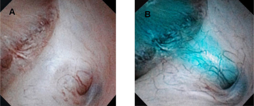
Figure 2 At the end of the procedure, a contrast study was performed and a double-J stent was retained inside the diverticulum. (A) Diverticular neck after widening. (B) Upper coil of double-J stent was in the diverticulum.
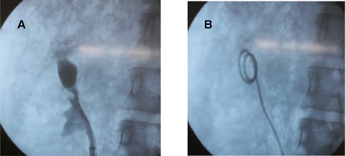
Table 1 Demographic Data
Table 2 Complications
Figure 3 The length of the diverticular neck was measured from the opening to the diverticulum.
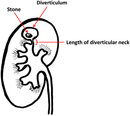
Figure 4 ROC curve, optimal cut-off point of stone size.
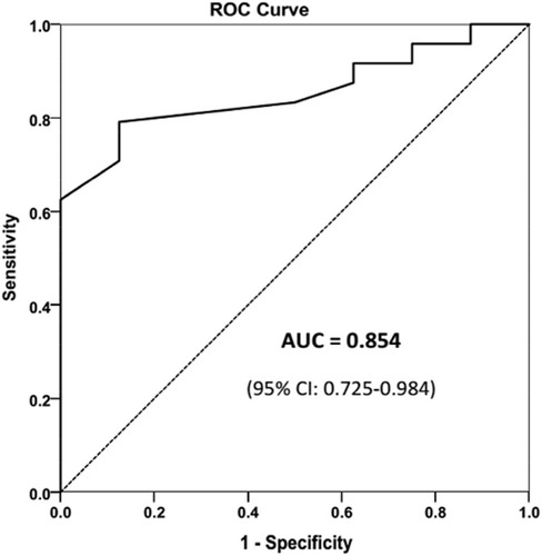
Figure 5 ROC curve, optimal cut-off point of diverticulum neck length.
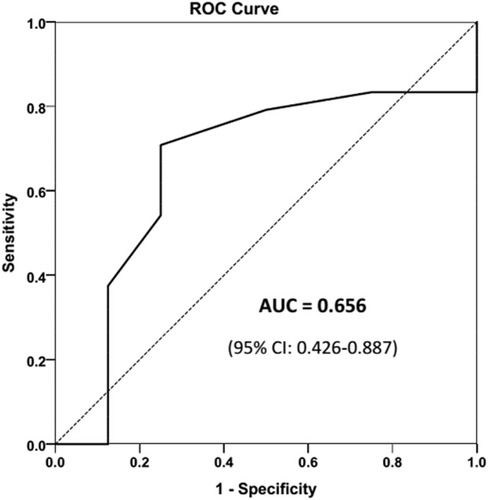
Table 3 Outcomes
Table 4 Multivariate Analysis of the Factors Affecting the Stone-Free Rate