Figures & data
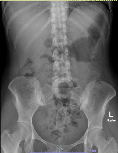
Figure 1 (A) KUB films at a gestational age of 4 weeks showing a left kidney stone that was approximately 3 cm in diameter. (B) Ultrasonography of the left kidney showing left hydronephrosis and a left kidney stone. (C) Ultrasonography of the bladder showing a normal bladder mucosa and a gestational sac in the uterus.
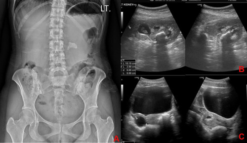
Figure 2 Endoscopic findings related to the infected kidney stone.
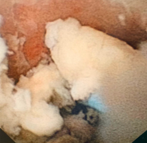
Figure 3 Timeline of kidney stone management.
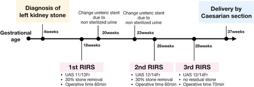
Figure 4 KUB films acquired 4 months after the final surgery.