Figures & data
Figure 1 Acne and facial hair (A) and enlarged penis with scant pubic hair (B) in a 6-year-old boy, showing signs of precocious pseudo-puberty as shown on both cases. Advanced bone age of Case 2 (C).
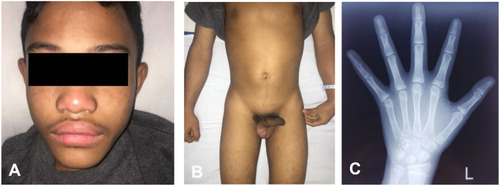
Table 1 Characteristics of Patients
Figure 2 Ultrasound of the testicles of Case 1 (A) and Case 2 (B).
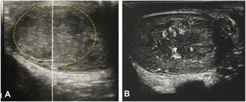
Table 2 Hormonal Assessment of the Patients
Figure 3 Macroscopic anatomy findings of the left testis from the patient of Case 1 (A) and Case 2 (B). Note a well circumscribed yellowish-brownish mass inside the testis.
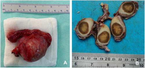
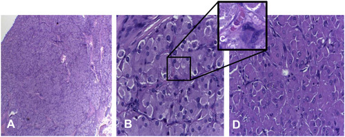
Figure 4 (A) The tumor mass was lobulated had indistinct border; (B) spherical nuclei of tumor cells, pleomorphic, granulated eosinophilic cytoplasm, (high power field, 400x); (C) Reinke crystal. (D) Solid tumors formed nest.