
Figures & data
Figure 1 Dextranomer/hyaluronic acid (Dx/HA) injection therapies for primary vesicoureteral reflux. *Extravesical portion of ureter (adjacent to ureteral hiatus). ×: position of needle insertion. (A) Double hydrodistention implantation technique (double HIT). (B) Systematic-multisite hydrodistention implantation technique (SMHIT) These figures show the coronal section of the ureterovesical junction. Multiple Dx/HA injections are sequentially and systematically administered from the proximal to the distal intramural ureter in SMHIT. The coaptation area of the intramural ureter created by Dx/HA bulges can be extended out further by SMHIT than by double HIT.
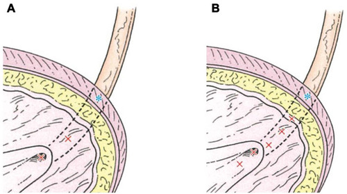
Figure 2 The first half of SMHIT (systematic-multisite hydrodistention implantation technique) for left VUR grade Ⅳ. *Extravesical portion of ureter (adjacent to ureteral hiatus). (A) Left ureteral orifice before SMHIT. (B) Mild opening of the ureteral orifice during hydrodistention. (C) intramural ureter. (D) First insertion of a needle and injection of Dx/HA. (E) Bulge after first injection of Dx/HA and second insertion of a needle. (F) Second injection of Dx/HA. (G) Bulge after second injection of Dx/HA. (H) Bulge after second injection of Dx/HA and third insertion of a needle.
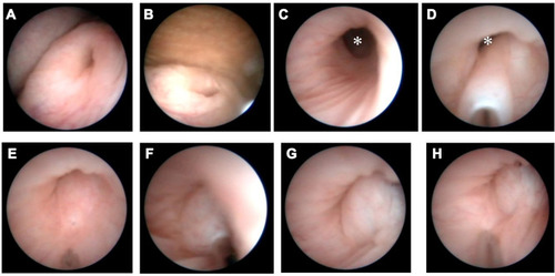
Figure 3 The latter half of SMHIT (systematic-multisite hydrodistention implantation technique) for left VUR grade Ⅳ (A and B) Third injection of Dx/HA. (C) Fourth insertion of a needle. (D and E) Fourth injection of Dx/HA (STING (subureteral transurethral injection) technique). (F) Fifth insertion of a needle. (G) Fifth injection of Dx/HA. (H) Left ureteral orifice after SMHIT.
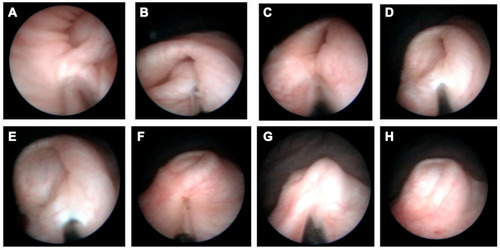
Table 1 Preoperative Patient Characteristics
Table 2 Intraoperative and Postoperative Results