
Figures & data
Figure 1 Axial CT images showing multiple abnormal nodular thickening on the anterior (A) and posterior (B) bladder walls.
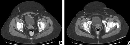
Figure 2 Bladder MALT lymphoma. The histologic sections show a dense, abnormal lymphocytic proliferation right beneath the urothelial surface of urinary bladder mucosa (A and B) the neoplastic cells are positive for CD20 (C) and CD79a (D).
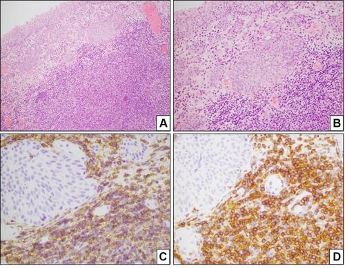
Figure 3 Axial CT image of depicting the bladder and showing no evidence of abnormal nodularity 3 months later.
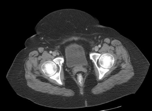
Table 1 Urinary Bladder MALT Lymphoma Cases: Diagnostics and Treatment