
Figures & data
Figure 1 Intraoperative picture; left polyorchidism with a separate epididymis and common vas deferens. Caudal testis (A), cranial testis (B), common vas (black arrow).
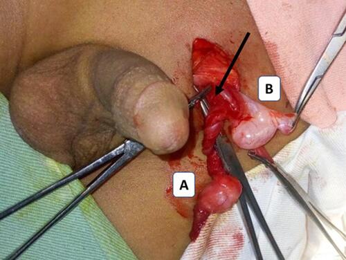
Table 1 Summary of the Literature Review of Case Reports and Case Series