Figures & data
Figure 1 Cross-sectional image of patient showed left renal vein (R) compressed between SMA (S) and aorta (A) with dilatation of left renal vein. Dilated right renal pelvis (p) can also be seen.
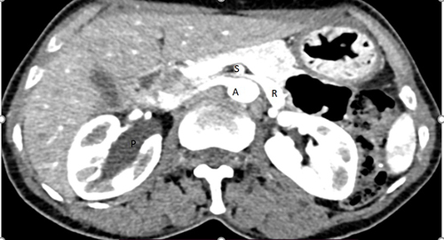
Figure 2 Cross-sectional image shows dilated left gonadal vein (g) going along side the aorta (a) and dilated right renal pelvis (p).
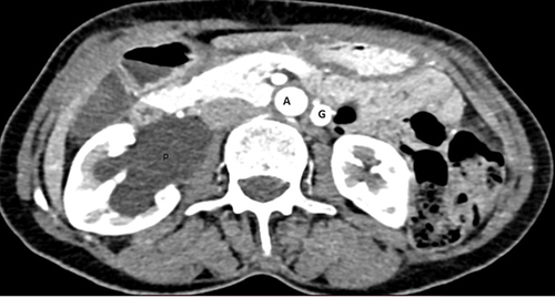
Figure 3 (A) coronal images showed dilated left gonadal vein (G). (B) right UPJO on delayed film with no passage of contrast to right ureter (P).
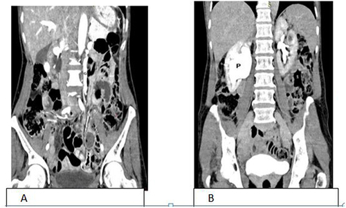
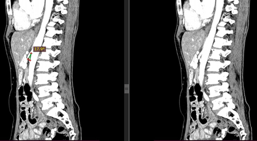
Figure 4 Sagittal view shows narrow angle between aorta and SMA, around 18° also slit of renal vein seen while passing between SMA and aorta.