Figures & data
Figure 1 CT scan findings.
Notes: (A) Contrast-enhanced CT scan shows a hyperdense rounded lesion, of 16 mm at its greatest diameter (arrow), in the inferior third of the right kidney, compatible with a posttraumatic pseudoaneurysm. (B) CT scan shows a 3 cm laceration and a perirenal hematoma in the posteroinferior third of the right kidney (arrow).
Abbreviation: CT, computed tomography.
Abbreviation: CT, computed tomography.
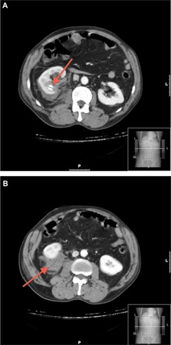
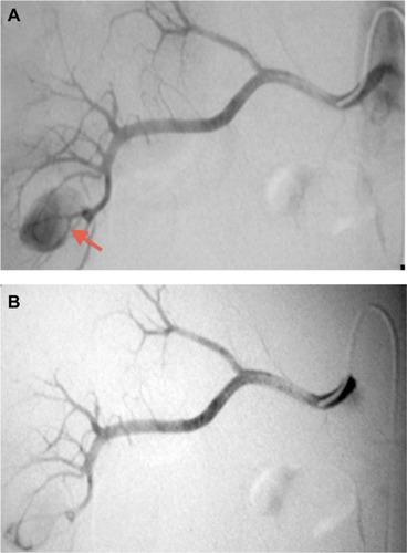
Figure 2 Right renal artery arteriogram findings.
Notes: (A) Ventral and dorsal branches of the right renal artery selective angiogram revealed a mesorenal pseudoaneurysm, of 16 mm at its greatest diameter (arrow), from two interlobar branches of the posterior division of the right renal artery. (B) Superselective embolization of two interlobar branches of the posterior division of the right renal artery was performed, and postembolization images confirmed complete pseudoaneurysm devascularization and preservation of the remaining renal vasculature.