Figures & data
Table 1 The primer sets used for amplification of studied genes by real-time polymerase chain reaction
Figure 1 Serum induces the upregulation of markers for glial differentiation. A) Western blot analysis for comparison of glial and neuronal markers in human mesenchymal stem cells cultured with or without 10% FBS. B) Densitometric measurement of Western blot protein bands (*P < 0.05). C) Immunofluorescence staining of Gal-C and GFAP expression in human mesenchymal stem cells cultured in different concentration of FBS. D) effects of serum deprivation on C6 rat glial cells expressing GFAP and Gal-C proteins.
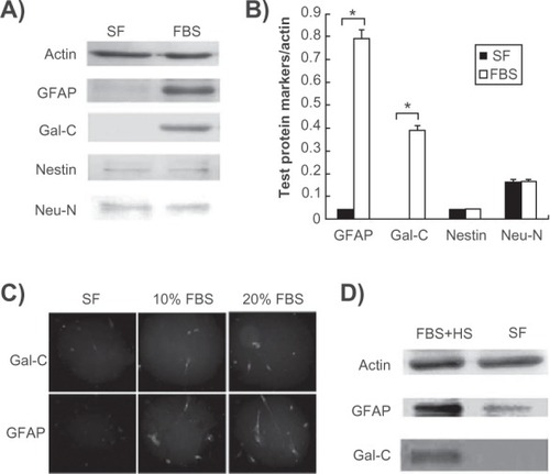
Figure 2 Recovery of glial markers by serum supplementation in serum-deprived human mesenchymal stem cells. A) Western blot analysis detecting the expression of Gal-C in human mesenchymal stem cells that were cultured in serum-free medium for two days and then supplemented with FBS. B) Immunofluorescence staining of glial fibrillary acidic protein expression in human mesenchymal stem cells cultured in either SF for two days or thereafter supplemented with 10% FBS for an additional two days (SF + FBS). C) Semiquantitative real-time polymerase chain reaction detecting mRNA levels of glial and oligodendrocyte markers using the above experimental conditions. C is a control that was continuously cultured in FBS-supplemented medium.
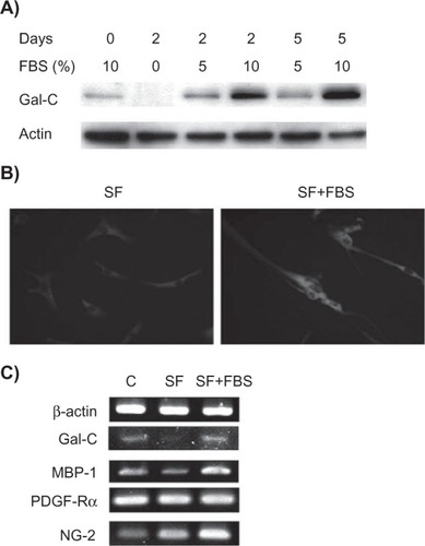
Figure 3 Effects of Notch signaling on serum-induced oligodendrocyte differentiation. A) Semiquantitative real-time polymerase chain reaction for detecting mRNA levels of JAK1 and Notch-associated markers in human mesenchymal stem cells cultured in different concentration of FBS. B) Western blotting for detecting the Gal-C and Notch1 cleaved internal domain in human mesenchymal stem cells treated with JAK1 inhibitor and Notch inhibitor MG132 for 24 hours. C) Comparison of effects of MG132 and lactacystin on expression of Gal-C. Upper panel: Western blot analysis. Lower panel: densitometric measurement of protein bands (*P < 0.05 compared with FBS control). D) Comparison of mRNA levels of oligodendrocyte-specific transcription factors (Olig-1, Olig-2, and SOX-10) in human mesenchymal stem cells treated with MG132 or lactacystin. E) Morphologic changes in GFP-labeled human mesenchymal stem cells. F) Comparison of the expression of MBP-1 in GFP-labeled human mesenchymal stem cells before (−) and after (+) MG132 treatment for 24 hours. Human mesenchymal stem cells were maintained in FBS during the treatment. The nuclei were stained by 4′,6-diamidino-2-phenylindole (DAPI). The sizes of scale bars for E and F were 20 and 40 μm, respectively.
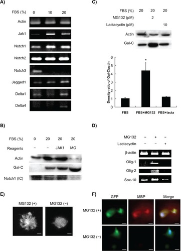
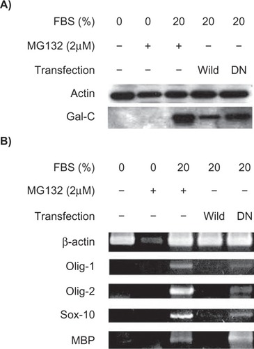
Figure 4 Inhibition of Notch activity by mutant RBP-J protein enhances the serum-stimulated expression of oligodendrocyte markers. A) Western blot analysis for comparison of Gal-C expression in human mesenchymal stem cells transfected with wild-type or the DN mutant form of RBP-J cDNA. B) Semiquantitative real-time polymerase chain reaction detecting the mRNA levels of other oligodendrocyte markers. MG132 (24 hours of treatment) was unable to induce these markers under serum-free conditions.