401
Views
0
CrossRef citations to date
0
Altmetric
REVIEW

Mitochondria in Cancer Stem Cells: From an Innocent Bystander to a Central Player in Therapy Resistance

ORCID Icon, ORCID Icon & ORCID Icon
Pages 19-41 | Received 23 Apr 2023, Accepted 15 Aug 2023, Published online: 23 Aug 2023

Figures & data

Table 1 Cancer Stem Cell Associated Markers Reported in Different Cancers

Figure 1 Mitochondrial Dysfunction and Cancer Stem Cells (CSCs). (a) Schematic of mitochondrial biogenesis and its regulation by transcription factors (PGC1, NRF, TFAM, PPAR, and ERR). Additionally, depicted are AMPK, oncogenic KRAS, and c-MYC-dependent mechanisms that lead to increase in biogenesis and energy production. This results in elevated oxidative phosphorylation (OXPHOS) and high ATP levels in CSCs. (b) Representation of mitochondrial metabolic dependency in CSCs. Cellular energy is derived through OXPHOS, fatty acid oxidation (FAO), and the TCA cycle within the mitochondria. CSCs exhibit increased oxidative phosphorylation for enhanced ATP production and elevated fatty acid oxidation through activation of oncogenic pathways. Deregulated TCA cycle enzymes in CSCs produce oncometabolites contributing to cancer progression. (c) Representation of altered mitochondrial dynamics in CSCs, where the balance between mitochondrial fission and fusion is disrupted. Upregulation of mitochondrial fission proteins (Drp1) and their regulators (Fis1, MID49, MID51, MFF) and downregulation of mitochondrial fusion proteins (Mfn1, Mfn2, OPA1) leads to impaired mitochondrial dynamics. (d) Increased activity of Ca2+-dependent kinases (PKC, CaN, CAMKIV, JNK, MAPK) due to altered membrane potential in CSCs is shown. Also indicated are the kinases and nuclear transcription factors involved in retrograde signaling. (e) Schematic representation of mitophagy in CSCs. Elevated cytoplasmic PINK1 phosphorylates Parkin and ubiquitinated-OMM proteins. Phosphorylated Parkin is transported into the mitochondria where it ubiquitinates itself and other mitochondrial substrates. These ubiquitin (Ub)-marked mitochondria are degraded by autophagosomes. (f) Mitochondria-mediated apoptosis in CSCs. Cells with damaged DNA activate caspase-8 mediated cell death. In CSCs, activation of caspase-8 is inhibited by high levels of c-FLIP; levels of pro-apoptotic proteins (Bax, Bak) are decreased while levels of anti-apoptotic proteins (Bcl-xL) are increased leading to cell survival and no apoptosis.

Figure 1 Mitochondrial Dysfunction and Cancer Stem Cells (CSCs). (a) Schematic of mitochondrial biogenesis and its regulation by transcription factors (PGC1, NRF, TFAM, PPAR, and ERR). Additionally, depicted are AMPK, oncogenic KRAS, and c-MYC-dependent mechanisms that lead to increase in biogenesis and energy production. This results in elevated oxidative phosphorylation (OXPHOS) and high ATP levels in CSCs. (b) Representation of mitochondrial metabolic dependency in CSCs. Cellular energy is derived through OXPHOS, fatty acid oxidation (FAO), and the TCA cycle within the mitochondria. CSCs exhibit increased oxidative phosphorylation for enhanced ATP production and elevated fatty acid oxidation through activation of oncogenic pathways. Deregulated TCA cycle enzymes in CSCs produce oncometabolites contributing to cancer progression. (c) Representation of altered mitochondrial dynamics in CSCs, where the balance between mitochondrial fission and fusion is disrupted. Upregulation of mitochondrial fission proteins (Drp1) and their regulators (Fis1, MID49, MID51, MFF) and downregulation of mitochondrial fusion proteins (Mfn1, Mfn2, OPA1) leads to impaired mitochondrial dynamics. (d) Increased activity of Ca2+-dependent kinases (PKC, CaN, CAMKIV, JNK, MAPK) due to altered membrane potential in CSCs is shown. Also indicated are the kinases and nuclear transcription factors involved in retrograde signaling. (e) Schematic representation of mitophagy in CSCs. Elevated cytoplasmic PINK1 phosphorylates Parkin and ubiquitinated-OMM proteins. Phosphorylated Parkin is transported into the mitochondria where it ubiquitinates itself and other mitochondrial substrates. These ubiquitin (Ub)-marked mitochondria are degraded by autophagosomes. (f) Mitochondria-mediated apoptosis in CSCs. Cells with damaged DNA activate caspase-8 mediated cell death. In CSCs, activation of caspase-8 is inhibited by high levels of c-FLIP; levels of pro-apoptotic proteins (Bax, Bak) are decreased while levels of anti-apoptotic proteins (Bcl-xL) are increased leading to cell survival and no apoptosis.

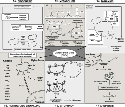
Table 2 Drug Category and Mode of Acquired Resistance in Cancer Stem Cells in Different Cancers