Figures & data
Table 1 Clinical effects of cell therapy for acute or chronic heart failure with different types of stem cells
Figure 1 Progression of stem cells in vitro into cardiomyocytes for in vivo transplantation.
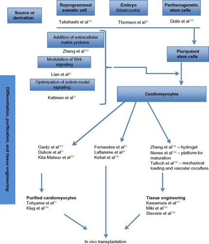
Figure 2 Clinical trials of cell therapy for acute and chronic, ischemic and nonischemic heart failure.
Abbreviations: ASTAMI, Autologous Stem-cell Transplantation in Acute Myocardial Infarction; BOOST, BOne marrOw transfer to enhance ST-elevation infarct regeneration; CADUCEUS, CArdiosphere-Derived aUtologous Stem CElls to reverse ventricUlar dySfunction; LateTIME, Use of Adult Autologous Stem Cells in Treating People 2 to 3 Weeks after having a Heart Attack; MYSTAR, MYocardial STem cell Administration after acute myocardial infaRction; POSEIDON, Comparison of Allogeneic versus Autologous Bone Marrow-Derived Mesenchymal Stem Cells Delivered by Trans-Endocardial Injection in Patients with Ischemic Cardiomyopathy; PRECISE, AdiPose-deRived stEm and Regenerative Cells In the Treatment of Patients with non revaScularizable ischEmic myocardium; REGENT, Myocardial REGENeraTion by intracoronary infusion of selected population of stem cells in acute myocardial infarction; REPAIR-AMI, Reinfusion of Enriched Progenitor cells And Infarct Remodeling in Acute Myocardial Infarction; SCIPIO, Stem Cell Infusion in Patients with Ischemic cardiOmyopathy; STAR-heart, The acute and long-term effects of intracoronary Stem cell Transplantation in patients with chronic heARt failure; SWISS-AMI, SWiss Multicenter Intracoronary Stem cells Study in Acute Myocardial Infarction; TOPCARE-AMI, Transplantation of Progenitor Cells and Regeneration Enhancement in Acute Myocardial Infarction; TOPCARE-CHD, Trans-Coronary Transplantation of Functionally Competent BMD Stem Cells; TOPCARE-DCM, Selective Intracoronary Bone Marrow-Derived Progenitor Cell Infusion in Patients with Non-Ischemic Dilated Cardiomyopathy.
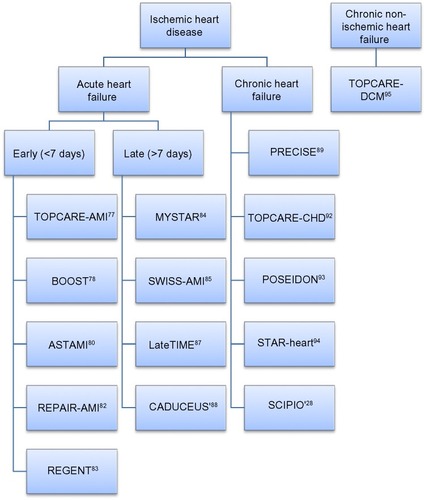
Table 2 Cells number and purification methods
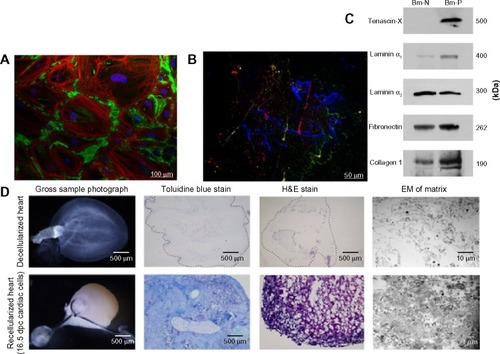
Figure 3 Biomimetic scaffolds
Notes: (A) Synthesis of biomatrix: fibroblasts isolated from samples of adult human heart were cultured in confluent state allowing for extracellular matrix (ECM) deposition in vitro. Representative image obtained by immunofluorescent labeling of actin filaments (red), cell nuclei (blue), and fibronectin (green). (B) Decellularization of biomatrix: after nonenzymatic removal of fibroblasts, ECM was observed under fluorescence microscope. Its composition was revealed by indirect immunofluorescent staining of representative ECM proteins: collagen IV (red), laminin (green), fibronectin (blue), and tenascin-C (yellow). (C) Immunoblotting of the decellularized biomatrix further confirmed the presence of these ECM-specific components in the biomatrix. Adapted from: Clotilde Castaldo, Franca Di Meglio, Rita Miraglia, et al., “Cardiac Fibroblast-Derived Extracellular Matrix (Biomatrix) as a Model for the Studies of Cardiac Primitive Cell Biological Properties in Normal and Pathological Adult Human Heart,” BioMed Research International, vol. 2013, Article ID 352370, 7 pages, 2013. doi:10.1155/2013/352370.Citation210 (D) Decellularization of embryonic cardiac tissue and recellularization with E16.5 ventricular cells. The constructs create a favorable microenvironment for the cells to integrate and migrate on the scaffold. Macroscopic appearance of the supporting matrix changed from translucent to opaque following cell inclusion into the construct. This was further confirmed by hematoxylin and eosin (H&E) and toluidine blue staining of the reseeded scaffolds, revealing a highly cellular environment on the host matrix. Collagen structures also became more physically compact after incubation of the scaffold with the cells, as shown by electron microscopy (EM). Adapted from Cree Chamberland, Almudena Martinez-Fernandez, Rosanna Beraldi, and Timothy J. Nelson, “Embryonic Decellularized Cardiac Scaffold Supports Embryonic Stem Cell Differentiation to Produce Beating Cardiac Tissue,” ISRN Stem Cells, vol. 2014, Article ID 625164, 10 pages, 2014. doi:10.1155/2014/625164.Citation211
Abbreviations: Bm-N, biomatrix from patient with a normal heart; Bm-P, biomatrix from patients with heart failure; dpc, day post conception.
Abbreviations: Bm-N, biomatrix from patient with a normal heart; Bm-P, biomatrix from patients with heart failure; dpc, day post conception.