Figures & data
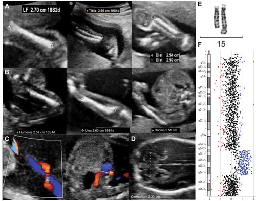
Figure 1 Morphologic ultrasonogram of a 21+1-week male fetus with 19+5-week-ultrasound parameters.
Notes: Bone morphology and mineralization characteristics were normal. (A) From left to right: FL 27-27 mm, tibia 25-26 mm, and fibula 25-25 mm. (B) From left to right: humerus 25-26 mm, ulna 25-26 mm, and radius 25-26 mm. (C) Single umbilical artery. (D) In Week 24, the transverse cerebellar diameter was 23 mm (<p5). Next, characterization of the duplication was carried out by cytogenetic and molecular analyses. (E) Conventional G-banding of chromosome 15. The duplicated chromosome is on the right. (F) CGH array. Reduced dosage for probes is shown to the left (red) of the control two-copy line and increased dosage is shown to the right (blue).
Abbreviation: CGH, comparative genomic hybridization.
Abbreviation: CGH, comparative genomic hybridization.