Figures & data
Table 1 Chromosomes and Genetic Defects Related to Moyamoya Angiopathy
Table 2 Molecular Biomarkers of Moyamoya Angiopathy by Protein Class
Figure 1 Potential disease mechanisms in MA. Red indicates that the factor affects vascular stenosis (intimal thickening, media attenuation, and internal elastic lamina damage). Blue indicates that the factor affects aberrant angiogenesis (capillary sprouting). Green indicates that the factor affects both vascular stenosis and aberrant angiogenesis.
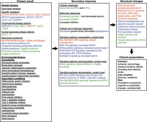
Figure 2 Proteins, cells, genes, and signaling pathways related to cerebral angiopathy characteristics in MA and associated disorders. (A) Arteriovenous malformation. (B) Intracranial aneurysm. (C) Vascular/internal carotid artery stenosis. (D) Stroke. Red indicates that the factor affects vascular stenosis (intimal thickening, media attenuation, and internal elastic lamina damage). Blue indicates that the factor affects aberrant angiogenesis (capillary sprouting). Green indicates that the factor affects both vascular stenosis and aberrant angiogenesis.
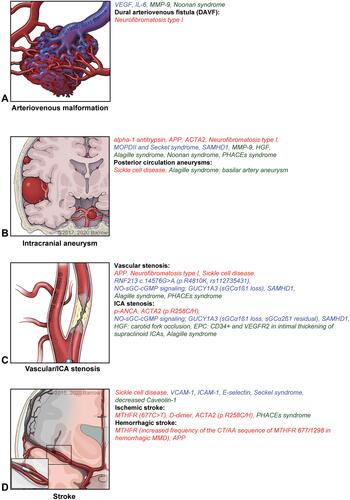
Figure 3 Schematic representation of an artery depicting macroscopic active sites of proteins, signaling pathways, genes, and cells in MA and associated disorders. Red indicates that the factor affects vascular stenosis (intimal thickening, media attenuation, and internal elastic lamina damage). Blue indicates that the factor affects aberrant angiogenesis (capillary sprouting). Green indicates that the factor affects both vascular stenosis and aberrant angiogenesis.
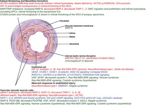
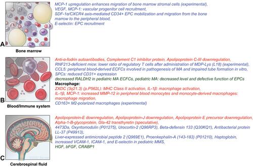
Figure 4 Microscopic active sites of proteins, cells, genes, and signaling pathways in MA and associated disorders. (A) Bone marrow. (B) Blood/immune system. (C) Cerebrospinal fluid. Red indicates that the factor affects vascular stenosis (intimal thickening, media attenuation, and internal elastic lamina damage). Blue indicates that the factor affects aberrant angiogenesis (capillary sprouting). Green indicates that the factor affects both vascular stenosis and aberrant angiogenesis.