Figures & data
Figure 1 Pedigrees of the four participating families. F1 refers to family #1; F2, family #2; F3, family #3, and F4 refers to family #4. Squares and circles indicate male and female members, respectively. Black filled symbols represent affected individuals of relevance in this study, red filled symbols represent affected siblings with different disease, empty symbols for unaffected individuals, crossed symbols for deceased individuals, double lines represent consanguinity, and finally, arrows refer to probands in each case.
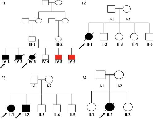
Table 1 Clinical Data for Participated Probands in Each Family
Table 2 Identified Variants in Each Participating Family
Table 3 Laboratory Tests for Both Patients in Family F1
Figure 2 Histopathological findings for patient IV-3 in family F1. (A and B) Hematoxylin and eosin staining shows steatosis, hepatocytes ballooning, and portal lymphocytic infiltration. (C and D) Masson’s trichrome and Van Gieson’s stain, respectfully, showing bridging fibrosis partially forming half nodules. Magnification: 40× (A and C) and 100× (B and D).
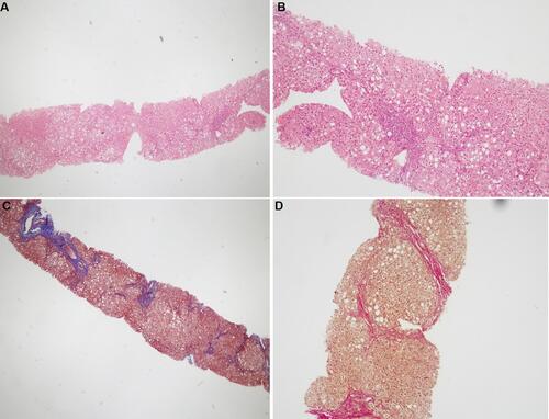
Figure 3 Sanger sequence chromatography for family F1 (left panel), family F2 (middle panel), and family F3 (right panel). In F1: the two probands (IV-1 and IV-3) have a pathogenic homozygous variant in SLC7A7 gene. Heterozygous carriers in the family are shown in middle part, while homozygous for the wild type allele is shown at the top. In F2 (middle panel): proband II-1 carries a de novo heterozygous variant in ACTG2 gene; whilst her parents carry the wild type homozygous allele. In the right panel, family F3, the two probands II-1 and II-2 have a homozygous deletion in HSD3B7 gene. While the parents along their children have the heterozygous allele.
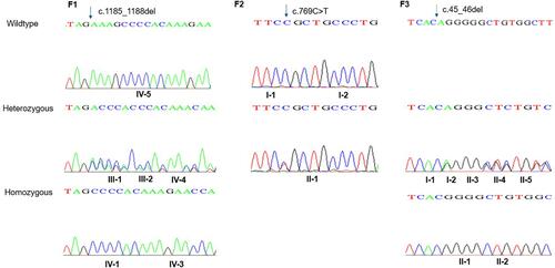
Figure 4 Imaging tests for proband II-1 in family F2. (A and B) Abdominal X-ray and barium study showing severe dilated bowel loops. (C and D) Calretinin staining 2 cm away from the anal verge, 4 cm from the anal verge (E), and from the ileostomy site (F). Ganglion cells are present (magnification: 40×, 100×, 200×, and 40× for C, D, E, and F, respectively).
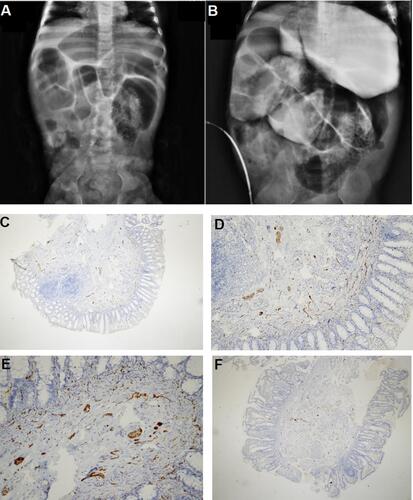
Table 4 Liver Function Tests and Blood Coagulation Tests Conforming Liver Damage in Proband II-1 from Family F3
Table 5 FAB-MS Test Results for Both Probands in Family F3 Indicate the Presence of Peaks Characteristic of 3-β-HSD Deficiency
Figure 5 Histopathological findings for patient II-1 in family F3. Hematoxylin and eosin staining showing hepatocytes ballooning and feathery changes with marked intrahepatic cholestasis. Mild periportal inflammation and foci of spotty necrosis are also present (magnification: 100× and 200× left to right, respectively).
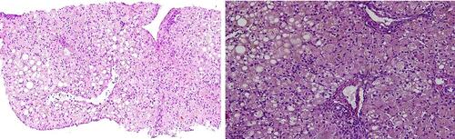
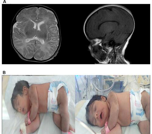
Figure 6 MRI and clinical features of proband II-2 in family F4. (A) Brain MRI; myelination is appropriate for proband’s age. Both thalami, both basal ganglia, corpus callosum, brainstem, cerebellum, and ventricular system appear normal. Central midline structures with no mass effect, no hemorrhage or collection seen. (B) Patient’s characteristic features; soft long ears, U-shaped vermilion of the upper lip, thick skin, hypertrichosis, moderate multiple contractures, hypotonia, and severe hypokinesia.