
Figures & data
Table 1 Age, and Weight and Height at Birth
Table 2 Clinical Variables of the Patients with DS
Figure 1 Dysmorphic phenotype in patient with DS. (A) Frontal view. (B) Smile in a frontal view. (C) Lateral view.
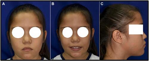
Figure 2 Dental photography. (A) Cross-bite. (B) The square in the medial incisors show enamel hypomineralization affecting both dentitions (C) superior maxillary (D) inferior maxillary, vertical overbite.
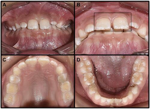
Figure 3 Dental photography (A) centric occlusion, it is evident the hyperpigmentation in the medial incisors and the generalize enamel hypoplasia. (B) Opening centric occlusion also shown the amalgam restoration of the lower molars, dental crowding in lower incisor and twisted of the upper first and second premolars.
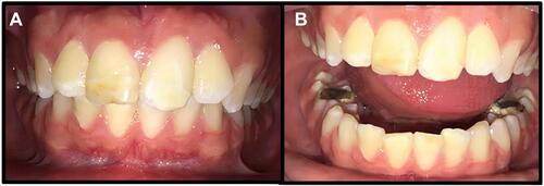
Figure 4 Panoramic dental X-ray. (A) Lateral view. (B) Panoramic dental X-ray shows dental crowding especially from the first and second superior premolars until the superior molars.
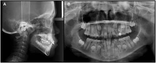
Table 3 Results of the Survey
Table 4 Summarizes the Current Literature About Di George Syndrome Dentistry Management and Craniofacial Characteristics