Figures & data
Figure 1 Cylindromas: germline and mosaic presentations. (A) Cylindromas and spiradenomas progressively grow and form a confluent mass, as seen in this severely affected patient with CCS. (B) Mosaic presentations of unilateral cylindromas, that may warrant skin biopsy and genetic testing to determine a recurrently detected pathogenic variant across multiple tumours. Image (B) reprinted from J Am Acad Dermatol, 81, Arefi M, Wilson V, Muthiah S et al. Diverse presentations of cutaneous mosaicism occur in CYLD cutaneous syndrome and may result in parent-to-child transmission. 1300–1307, Copyright (2019), with permission from Elsevier.Citation24
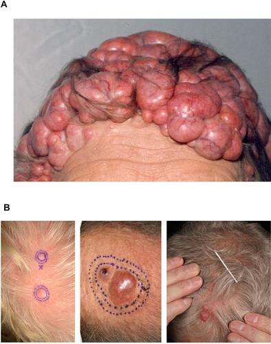
Figure 2 Skin tumours frequently seen in CCS. (A) Cylindroma. (B) Spiradenoma indicated by black arrow. (C) Trichoepithelioma indicated by white arrows. Reprinted from Dermatol Clin, 35 (1), Dubois A, Hodgson K, Rajan N. Understanding Inherited Cylindromas: Clinical Implications of Gene Discovery. 61–71, Copyright (2017), with permission from Elsevier.Citation116
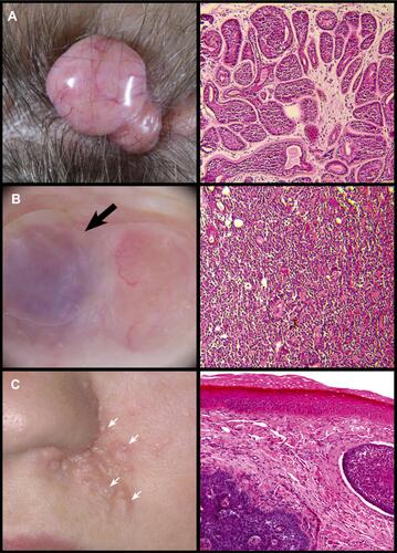
Figure 3 Pulmonary and cutaneous cylindromas visualised radiologically and endoscopically. (A) Spatial location of cutaneous CCS tumours seen on a CT with contrast indicated in green, and pulmonary CCS tumours indicated in yellow. Adapted from (B) Intra bronchial CCS tumour visualised during bronchoscopy. Adapted with permission from Brown SM, Arefi M, Stones R, et al. Inherited pulmonary cylindromas: extending the phenotype of CYLD mutation carriers. Br J Dermatol. 2018;179:662–668. © 2018 The Authors. British Journal of Dermatology published by John Wiley & Sons Ltd on behalf of British Association of Dermatologists.Citation41
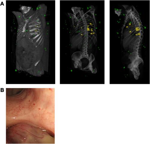
Figure 4 CYLD gene pathogenic variants identified to date. (A) Exonic locations of CYLD pathogenic variants in CCS patients; note a predisposition to the 3’ end of the gene, from which the catalytic domains are encoded. (B) Frameshift and nonsense pathogenic variants resulting in a predicted truncating protein are the most frequent mutation type seen.
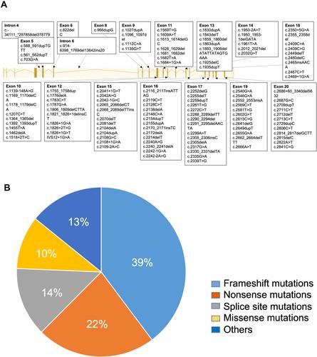
Table 1 List of Published Pathogenic Variants in CCS
Figure 5 A suggested testing strategy for CCS that addresses germline and mosaic presentations. *RNA from blood leucocytes require special collection tubes; **in principle such a patient may still yet harbour a germline pathogenic variant, and it remains the clinician’s decision if she would prefer to pursue GERMLINE testing first.
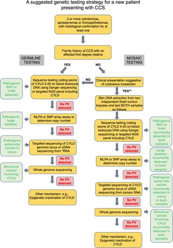
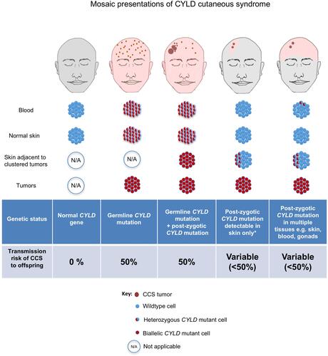
Figure 6 An overview of risk of transmission of germline and mosaic variants in CCS.