
Figures & data
Figure 1 Sanger sequencing results: electropherograms of the affected patient and her parents. (+) indicated the wild type allele. The position of the mutation is indicated by the arrow.
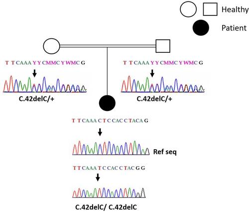
Figure 2 Sagittal T2-weighted MRI without injection showing bilobular ovarian mass (arrow) and ascites (asterisk).
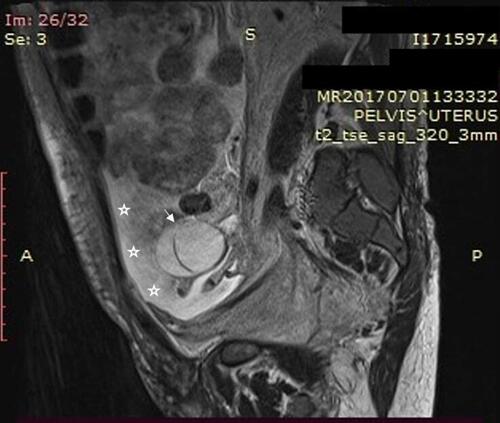
Figure 3 Serous cystadenoma of the ovary. (A) Thin-walled unilocular cyst that is lined by cubo-cylindrical monostratified epithelium (Low magnification x 10). (B) Monostratified, focally pseudostratified lining with monotonous, cuboidal or columnar, ciliated cells with round or oval nuclei. The epithelium is supported by variable amounts of spindle cell stroma with no cytologic atypia seen (Medium magnification x 20).
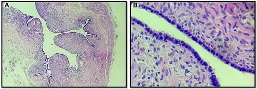
Table 1 Reported Clinical and Biological Findings Characterizing H Syndrome in the Literature Compared with Those in Our Patient