Figures & data
Figure 1 Structure of ClC-1 and localization of the mutation (p.R377X) in two Colombian siblings.
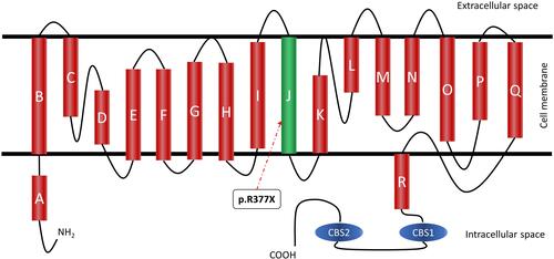
Table 1 Clinical Features of Two Patients with the CLCN1 Mutation
Figure 2 Familial pedigree.
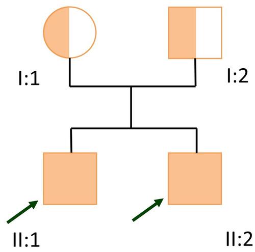
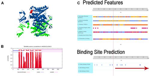
Figure 3 Effect of the Arg377* de novo mutation in the CLCN1 protein. (A) Figure shows the tridimensional structure of the CLCN1 protein chain A is in green and chain B in blue in the red dock is marked the c.1129C>T (p.Arg377deletion) CLCN1. (B) Graph B shows the graphical output of the Transmembrane Topology and Signal Peptide Prediction Using Dynamic Bayesian Networks (TMHMM) posterior probabilities sp P35523 CLCN1_Human, demonstrating the posterior probabilities for transmembrane (red), inside (blue), and outside (pink). In the case of the Chloride Voltage-Gated Channel 1 (CLCN1) six transmembrane domains are affected, three and two inside interactions, respectively are disrupted. (C) Predicted features the protein, which are disrupted for the early termination of the protein. The figure shows the predicted changes in the secondary structure, topology, DNA and RNA binding, and the binding site predicted disruption. The red line represents the initiation of the disruption in the protein done by the variant.