Figures & data
Figure 1 Cohesin complex and cohesin regulators.
Notes: Ring-embraced model of cohesion. The cohesin complex is formed by four proteins, ie, SMC1, SMC3, SCC1/RAD21, and SCC3/STAG. The SMC1, SMC3, and RAD21/SCC1 subunits form a ring-like structure. RAD21/SCC1 is the subunit that closes the ring formed by SMC (structural maintenance of chromosomes) subunits and has been named α-kleisin (closure). The SCC3/STAG protein interacts with RAD21/SCC1 to complete the cohesin complex. The adherin loading complex consists of two proteins, ie, SCC2/NIPBL and MAU2/SCC4. The acetyltransferases (ESCO1/2) acetylate the SMC3 cohesin subunit to establish cohesion of the chromatids. The cofactors, PDS5, wAPL, and Sororin, are involved in association and/or dissociation with chromatin.
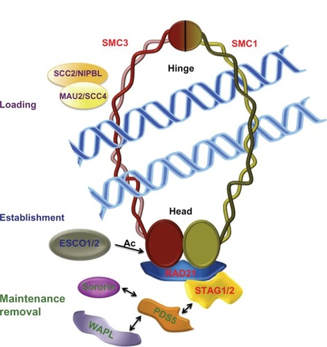
Table 1 Subunits of cohesin complexes and cohesin regulators implicated in cohesinopathies
Figure 2 Cohesin and cohesin regulators in human cohesinopathies.
Notes: Gene mutation occurs in human CdLS and RBS (orange arrows), and results in mouse models suggest involvement of cohesin and cohesin regulator genes in these human disorders (green arrows). Mutations in the SCC2/NIPBL gene cause the most severe phenotype in patients with Cornelia de Lange syndrome. Mutations in genes encoding for the cohesin subunits SMC1α, SMC3, and RAD21, and for the histone deacetylase, HDAC8, cause a mild variant of this syndrome. Although the phenotype of mice lacking PDS5A and/or PDS5B function show developmental abnormalities like those in patients with Cornelia de Lange syndrome, currently there are no data supporting mutations of these genes as a cause of this cohesinopathy in humans. Similarly, to the author’s knowledge, no data have been reported on STAG1 mutations in patients with Cornelia de Lange syndrome. A human ortholog of yeast Eco1 named ESCO2 is mutated in Roberts syndrome, which has a phenotype closely related to that seen in Cornelia de Lange syndrome.
Abbreviation: Ac, Acetylation.
Abbreviation: Ac, Acetylation.
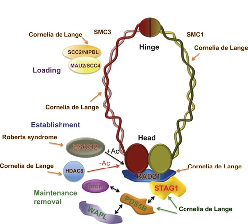
Figure 3 Models for a chromatin architectural function of cohesin in control of gene expression. (A) Interaction of the cohesin complex with chromatin-bound CTCF maintains a chromatin structure in which the enhancer cannot interact with the promoter and behaves as an insulator, repressing gene expression. (B) Loop structure formed by the mediator/cohesin complex allows enhancer-promoter interactions promoting gene transcription.
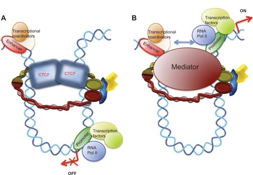
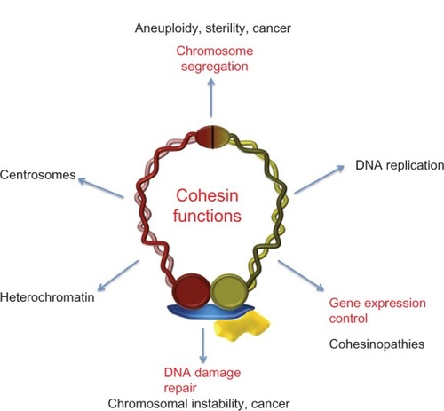
Figure 4 Cohesin metabolism and human disease.
Notes: Scheme of the main life processes of the cell in which cohesins have been functionally characterized and related diseases. The role of cohesin in control of gene expression (in red) is the principal focus of this review.